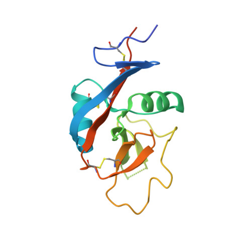
4J6K

Crystal structure of calcium2+-free wild-type CD23 lectin domain (crystal form B)


Experimental Data Snapshot

  • Method: X-RAY DIFFRACTION
  • Resolution: 2.30 Å
  • R-Value Free: 0.218 
  • R-Value Work: 0.191 
  • R-Value Observed: 0.193 

wwPDB Validation   3D Report Full Report


This is version 1.0 of the entry. See complete history


Literature

Conformational plasticity at the IgE-binding site of the B-cell receptor CD23.

Dhaliwal, B.Pang, M.O.Yuan, D.Yahya, N.Fabiane, S.M.McDonnell, J.M.Gould, H.J.Beavil, A.J.Sutton, B.J.

(2013) Mol Immunol 56: 693-697

  • DOI: https://doi.org/10.1016/j.molimm.2013.07.005
  • Primary Citation of Related Structures:  
    4J6J, 4J6K, 4J6L, 4J6M, 4J6N, 4J6P, 4J6Q

  • PubMed Abstract: 

    IgE antibodies play a central role in allergic disease. They recognize allergens via their Fab regions, whilst their effector functions are controlled through interactions of the Fc region with two principal cell surface receptors, FcɛRI and CD23. Crosslinking of FcɛRI-bound IgE on mast cells and basophils by allergen initiates an immediate inflammatory response, while the interaction of IgE with CD23 on B-cells regulates IgE production. We have determined the structures of the C-type lectin "head" domain of CD23 from seven crystal forms. The thirty-five independent structures reveal extensive conformational plasticity in two loops that are critical for IgE binding.


  • Organizational Affiliation

    King's College London, Randall Division of Cell and Molecular Biophysics, New Hunt's House, Guy's Campus, London SE1 1UL, UK; MRC & Asthma UK Centre in Allergic Mechanisms of Asthma, London, UK. Electronic address: bal.dhaliwal@kcl.ac.uk.


Macromolecules
Find similar proteins by:  (by identity cutoff)  |  3D Structure
Entity ID: 1
MoleculeChains Sequence LengthOrganismDetailsImage
Low affinity immunoglobulin epsilon Fc receptor
A, B, C, D, E
A, B, C, D, E, F, G, H
143Homo sapiensMutation(s): 0 
Gene Names: CD23ACLEC4JFCE2FCER2IGEBF
UniProt & NIH Common Fund Data Resources
Find proteins for P06734 (Homo sapiens)
Explore P06734 
Go to UniProtKB:  P06734
PHAROS:  P06734
GTEx:  ENSG00000104921 
Entity Groups  
Sequence Clusters30% Identity50% Identity70% Identity90% Identity95% Identity100% Identity
UniProt GroupP06734
Sequence Annotations
Expand
  • Reference Sequence
Experimental Data & Validation

Experimental Data

  • Method: X-RAY DIFFRACTION
  • Resolution: 2.30 Å
  • R-Value Free: 0.218 
  • R-Value Work: 0.191 
  • R-Value Observed: 0.193 
  • Space Group: P 1
Unit Cell:
Length ( Å )Angle ( ˚ )
a = 52.56α = 79.79
b = 56.13β = 84.85
c = 108.52γ = 70.22
Software Package:
Software NamePurpose
MOSFLMdata reduction
SCALAdata scaling
PHASERphasing
BUSTER-TNTrefinement
PDB_EXTRACTdata extraction
GDAdata collection
BUSTERrefinement

Structure Validation

View Full Validation Report


Entry History 

Deposition Data

Revision History  (Full details and data files)

  • Version 1.0: 2013-08-28
    Type: Initial release