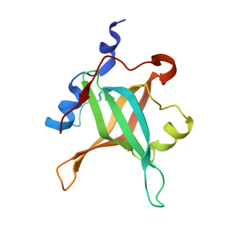
4IJL

Fragment-based Discovery of Protein-Protein Interaction Inhibitors of Replication Protein A


Experimental Data Snapshot

  • Method: X-RAY DIFFRACTION
  • Resolution: 1.70 Å
  • R-Value Free: 0.233 
  • R-Value Work: 0.191 
  • R-Value Observed: 0.193 

Starting Model: experimental
View more details

wwPDB Validation   3D Report Full Report


Ligand Structure Quality Assessment 


This is version 1.1 of the entry. See complete history


Literature

Discovery of Protein-Protein Interaction Inhibitors of Replication Protein A.

Patrone, J.D.Kennedy, J.P.Frank, A.O.Feldkamp, M.D.Vangamudi, B.Pelz, N.F.Rossanese, O.W.Waterson, A.G.Chazin, W.J.Fesik, S.W.

(2013) ACS Med Chem Lett 4: 601-605

  • DOI: https://doi.org/10.1021/ml400032y
  • Primary Citation of Related Structures:  
    4IJH, 4IJL

  • PubMed Abstract: 

    Replication Protein A (RPA) is a ssDNA binding protein that is essential for DNA replication and repair. The initiation of the DNA damage response by RPA is mediated by protein-protein interactions involving the N-terminal domain of the 70 kDa subunit with partner proteins. Inhibition of these interactions increases sensitivity towards DNA damage and replication stress and may therefore be a potential strategy for cancer drug discovery. Towards this end, we have discovered two lead series of compounds, derived from hits obtained from a fragment-based screen, that bind to RPA70N with low micromolar affinity and inhibit the binding of an ATRIP-derived peptide to RPA. These compounds may offer a promising starting point for the discovery of clinically useful RPA inhibitors.


  • Organizational Affiliation

    Department of Biochemistry, Vanderbilt University School of Medicine, Nashville, TN 37232 (USA).


Macromolecules
Find similar proteins by:  (by identity cutoff)  |  3D Structure
Entity ID: 1
MoleculeChains Sequence LengthOrganismDetailsImage
Replication protein A 70 kDa DNA-binding subunit123Homo sapiensMutation(s): 1 
Gene Names: REPA1RPA1RPA70
UniProt & NIH Common Fund Data Resources
Find proteins for P27694 (Homo sapiens)
Explore P27694 
Go to UniProtKB:  P27694
PHAROS:  P27694
GTEx:  ENSG00000132383 
Entity Groups  
Sequence Clusters30% Identity50% Identity70% Identity90% Identity95% Identity100% Identity
UniProt GroupP27694
Sequence Annotations
Expand
  • Reference Sequence
Small Molecules
Ligands 1 Unique
IDChains Name / Formula / InChI Key2D Diagram3D Interactions
1EK
Query on 1EK

Download Ideal Coordinates CCD File 
B [auth A],
C [auth A]
{[5-(3-chloro-1-benzothiophen-2-yl)-4-phenyl-4H-1,2,4-triazol-3-yl]sulfanyl}acetic acid
C18 H12 Cl N3 O2 S2
GNEUDTMFXMASKC-UHFFFAOYSA-N
Binding Affinity Annotations 
IDSourceBinding Affinity
1EK PDBBind:  4IJL Kd: 1.35e+5 (nM) from 1 assay(s)
BindingDB:  4IJL Kd: 1.35e+5 (nM) from 1 assay(s)
Experimental Data & Validation

Experimental Data

  • Method: X-RAY DIFFRACTION
  • Resolution: 1.70 Å
  • R-Value Free: 0.233 
  • R-Value Work: 0.191 
  • R-Value Observed: 0.193 
  • Space Group: P 21 21 21
Unit Cell:
Length ( Å )Angle ( ˚ )
a = 37.551α = 90
b = 53.569β = 90
c = 54.259γ = 90
Software Package:
Software NamePurpose
HKL-2000data collection
PHENIXmodel building
PHENIXrefinement
HKL-2000data reduction
HKL-2000data scaling
PHENIXphasing

Structure Validation

View Full Validation Report


Ligand Structure Quality Assessment 


Entry History 

Deposition Data

Revision History  (Full details and data files)

  • Version 1.0: 2013-08-14
    Type: Initial release
  • Version 1.1: 2023-09-20
    Changes: Data collection, Database references, Derived calculations, Refinement description