Discovery of Protein-Protein Interaction Inhibitors of Replication Protein A.
Patrone, J.D., Kennedy, J.P., Frank, A.O., Feldkamp, M.D., Vangamudi, B., Pelz, N.F., Rossanese, O.W., Waterson, A.G., Chazin, W.J., Fesik, S.W.(2013) ACS Med Chem Lett 4: 601-605
- PubMed: 23914285 Search on PubMedSearch on PubMed Central
- DOI: https://doi.org/10.1021/ml400032y
- Primary Citation Related Structures:
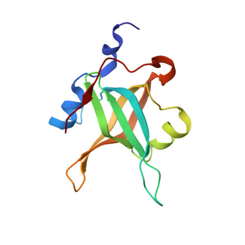
4IJH, 4IJL - PubMed Abstract:
Replication Protein A (RPA) is a ssDNA binding protein that is essential for DNA replication and repair. The initiation of the DNA damage response by RPA is mediated by protein-protein interactions involving the N-terminal domain of the 70 kDa subunit with partner proteins. Inhibition of these interactions increases sensitivity towards DNA damage and replication stress and may therefore be a potential strategy for cancer drug discovery. Towards this end, we have discovered two lead series of compounds, derived from hits obtained from a fragment-based screen, that bind to RPA70N with low micromolar affinity and inhibit the binding of an ATRIP-derived peptide to RPA. These compounds may offer a promising starting point for the discovery of clinically useful RPA inhibitors.
- Department of Biochemistry, Vanderbilt University School of Medicine, Nashville, TN 37232 (USA).
Organizational Affiliation: