4IHU | pdb_00004ihu

Reduced form of disulfide bond oxdioreductase (DsbG) from Mycobacterium tuberculosis


Experimental Data Snapshot

  • Method: X-RAY DIFFRACTION
  • Resolution: 1.90 Å
  • R-Value Free: 
    0.231 (Depositor), 0.231 (DCC) 
  • R-Value Work: 
    0.201 (Depositor), 0.203 (DCC) 
  • R-Value Observed: 
    0.202 (Depositor) 

Starting Model: experimental
View more details

wwPDB Validation 3D Report Full Report

Validation slider image for 4IHU

This is version 1.3 of the entry. See complete history

Literature

Structural and biochemical characterization of the essential DsbA-like disulfide bond forming protein from Mycobacterium tuberculosis.

Chim, N.Harmston, C.A.Guzman, D.J.Goulding, C.W.

(2013) BMC Struct Biol 13: 23-23

  • DOI: https://doi.org/10.1186/1472-6807-13-23
  • Primary Citation Related Structures: 
    4IHU

  • PubMed Abstract: 

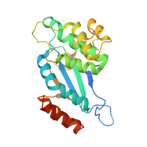
    Bacterial Disulfide bond forming (Dsb) proteins facilitate proper folding and disulfide bond formation of periplasmic and secreted proteins. Previously, we have shown that Mycobacterium tuberculosis Mt-DsbE and Mt-DsbF aid in vitro oxidative folding of proteins. The M. tuberculosis proteome contains another predicted membrane-tethered Dsb protein, Mt-DsbA, which is encoded by an essential gene. Herein, we present structural and biochemical analyses of Mt-DsbA. The X-ray crystal structure of Mt-DsbA reveals a two-domain structure, comprising a canonical thioredoxin domain with the conserved CXXC active site cysteines in their reduced form, and an inserted α-helical domain containing a structural disulfide bond. The overall fold of Mt-DsbA resembles that of other DsbA-like proteins and not Mt-DsbE or Mt-DsbF. Biochemical characterization demonstrates that, unlike Mt-DsbE and Mt-DsbF, Mt-DsbA is unable to oxidatively fold reduced, denatured hirudin. Moreover, on the substrates tested in this study, Mt-DsbA has disulfide bond isomerase activity contrary to Mt-DsbE and Mt-DsbF. These results suggest that Mt-DsbA acts upon a distinct subset of substrates as compared to Mt-DsbE and Mt-DsbF. One could speculate that Mt-DsbE and Mt-DsbF are functionally redundant whereas Mt-DsbA is not, offering an explanation for the essentiality of Mt-DsbA in M. tuberculosis.


  • Organizational Affiliation
    • Departments of Molecular Biology and Biochemistry, UCI, Irvine, CA 92697, USA. celia.goulding@uci.edu.

Macromolecule Content 

  • Total Structure Weight: 47.29 kDa 
  • Atom Count: 3,307 
  • Modeled Residue Count: 409 
  • Deposited Residue Count: 448 
  • Unique protein chains: 1

Macromolecules

Find similar proteins by:|  3D Structure
Entity ID: 1
MoleculeChains  Sequence LengthOrganismDetailsImage
Isomerase DsbG
A, B
224Mycobacterium tuberculosis H37RvMutation(s): 0 
Gene Names: Rv2969c
UniProt
Find proteins for O33272 (Mycobacterium tuberculosis (strain ATCC 25618 / H37Rv))
Explore O33272 
Go to UniProtKB:  O33272
Entity Groups
Sequence Clusters30% Identity50% Identity70% Identity90% Identity95% Identity100% Identity
UniProt GroupO33272
Sequence Annotations
Expand
Reference Sequence

Small Molecules

Ligands 1 Unique
IDChains Name / Formula / InChI Key2D Diagram3D Interactions
CL

Query on CL


Download:Ideal Coordinates CCD File
C [auth A]CHLORIDE ION
Cl
VEXZGXHMUGYJMC-UHFFFAOYSA-M
Modified Residues  1 Unique
IDChains TypeFormula2D DiagramParent
MSE
Query on MSE
A, B
L-PEPTIDE LINKINGC5 H11 N O2 SeMET

Experimental Data & Validation

Experimental Data

  • Method: X-RAY DIFFRACTION
  • Resolution: 1.90 Å
  • R-Value Free:  0.231 (Depositor), 0.231 (DCC) 
  • R-Value Work:  0.201 (Depositor), 0.203 (DCC) 
  • R-Value Observed: 0.202 (Depositor) 
Space Group: P 21 21 2
Unit Cell:
Length ( Å )Angle ( ˚ )
a = 71.03α = 90
b = 76.71β = 90
c = 86.96γ = 90
Software Package:
Software NamePurpose
ADSCdata collection
SHELXSphasing
PHENIXrefinement
MOSFLMdata reduction
SCALAdata scaling

Structure Validation

View Full Validation Report


Entry History 

Deposition Data

Revision History  (Full details and data files)

  • Version 1.0: 2013-10-30
    Type: Initial release
  • Version 1.1: 2023-09-20
    Changes: Data collection, Database references, Derived calculations, Refinement description
  • Version 1.2: 2023-12-06
    Changes: Data collection
  • Version 1.3: 2024-11-20
    Changes: Structure summary