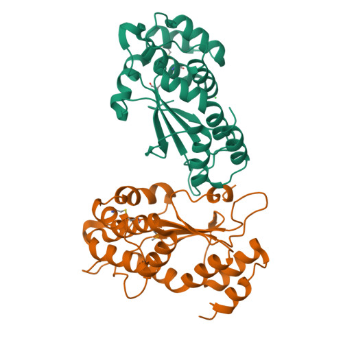
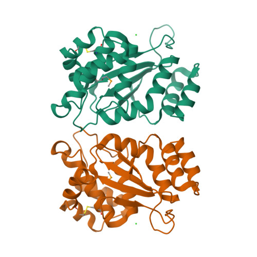
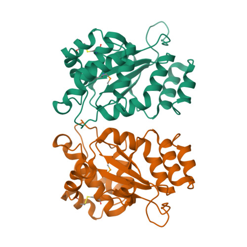
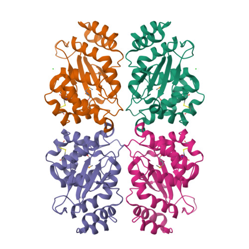
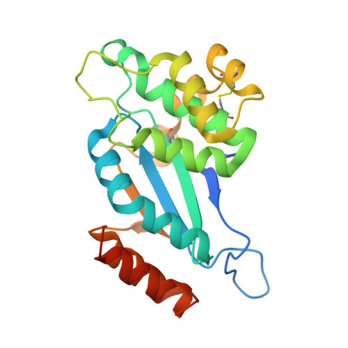
4IHU

Reduced form of disulfide bond oxdioreductase (DsbG) from Mycobacterium tuberculosis


Experimental Data Snapshot

  • Method: X-RAY DIFFRACTION
  • Resolution: 1.90 Å
  • R-Value Free: 0.231 
  • R-Value Work: 0.201 
  • R-Value Observed: 0.202 

Starting Model: experimental
View more details

wwPDB Validation   3D Report Full Report


This is version 1.3 of the entry. See complete history


Literature

Structural and biochemical characterization of the essential DsbA-like disulfide bond forming protein from Mycobacterium tuberculosis.

Chim, N.Harmston, C.A.Guzman, D.J.Goulding, C.W.

(2013) BMC Struct Biol 13: 23-23

  • DOI: https://doi.org/10.1186/1472-6807-13-23
  • Primary Citation of Related Structures:  
    4IHU

  • PubMed Abstract: 

    Bacterial Disulfide bond forming (Dsb) proteins facilitate proper folding and disulfide bond formation of periplasmic and secreted proteins. Previously, we have shown that Mycobacterium tuberculosis Mt-DsbE and Mt-DsbF aid in vitro oxidative folding of proteins. The M. tuberculosis proteome contains another predicted membrane-tethered Dsb protein, Mt-DsbA, which is encoded by an essential gene. Herein, we present structural and biochemical analyses of Mt-DsbA. The X-ray crystal structure of Mt-DsbA reveals a two-domain structure, comprising a canonical thioredoxin domain with the conserved CXXC active site cysteines in their reduced form, and an inserted α-helical domain containing a structural disulfide bond. The overall fold of Mt-DsbA resembles that of other DsbA-like proteins and not Mt-DsbE or Mt-DsbF. Biochemical characterization demonstrates that, unlike Mt-DsbE and Mt-DsbF, Mt-DsbA is unable to oxidatively fold reduced, denatured hirudin. Moreover, on the substrates tested in this study, Mt-DsbA has disulfide bond isomerase activity contrary to Mt-DsbE and Mt-DsbF. These results suggest that Mt-DsbA acts upon a distinct subset of substrates as compared to Mt-DsbE and Mt-DsbF. One could speculate that Mt-DsbE and Mt-DsbF are functionally redundant whereas Mt-DsbA is not, offering an explanation for the essentiality of Mt-DsbA in M. tuberculosis.


  • Organizational Affiliation

    Departments of Molecular Biology and Biochemistry, UCI, Irvine, CA 92697, USA. celia.goulding@uci.edu.


Macromolecules
Find similar proteins by:  (by identity cutoff)  |  3D Structure
Entity ID: 1
MoleculeChains Sequence LengthOrganismDetailsImage
Isomerase DsbG
A, B
224Mycobacterium tuberculosis H37RvMutation(s): 0 
Gene Names: Rv2969c
UniProt
Find proteins for O33272 (Mycobacterium tuberculosis (strain ATCC 25618 / H37Rv))
Explore O33272 
Go to UniProtKB:  O33272
Entity Groups  
Sequence Clusters30% Identity50% Identity70% Identity90% Identity95% Identity100% Identity
UniProt GroupO33272
Sequence Annotations
Expand
  • Reference Sequence
Small Molecules
Ligands 1 Unique
IDChains Name / Formula / InChI Key2D Diagram3D Interactions
CL
Query on CL

Download Ideal Coordinates CCD File 
C [auth A]CHLORIDE ION
Cl
VEXZGXHMUGYJMC-UHFFFAOYSA-M
Modified Residues  1 Unique
IDChains TypeFormula2D DiagramParent
MSE
Query on MSE
A, B
L-PEPTIDE LINKINGC5 H11 N O2 SeMET
Experimental Data & Validation

Experimental Data

  • Method: X-RAY DIFFRACTION
  • Resolution: 1.90 Å
  • R-Value Free: 0.231 
  • R-Value Work: 0.201 
  • R-Value Observed: 0.202 
  • Space Group: P 21 21 2
Unit Cell:
Length ( Å )Angle ( ˚ )
a = 71.03α = 90
b = 76.71β = 90
c = 86.96γ = 90
Software Package:
Software NamePurpose
ADSCdata collection
SHELXSphasing
PHENIXrefinement
MOSFLMdata reduction
SCALAdata scaling

Structure Validation

View Full Validation Report


Entry History 

Deposition Data

Revision History  (Full details and data files)

  • Version 1.0: 2013-10-30
    Type: Initial release
  • Version 1.1: 2023-09-20
    Changes: Data collection, Database references, Derived calculations, Refinement description
  • Version 1.2: 2023-12-06
    Changes: Data collection
  • Version 1.3: 2024-11-20
    Changes: Structure summary