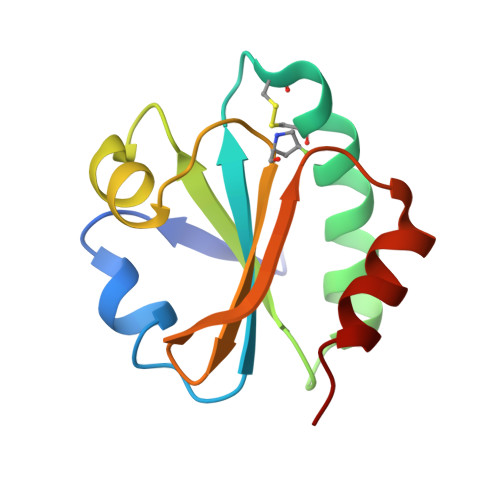
4HU9

E. coli thioredoxin variant with (4S)-FluoroPro76 as single proline residue


Experimental Data Snapshot

  • Method: X-RAY DIFFRACTION
  • Resolution: 1.55 Å
  • R-Value Free: 0.210 
  • R-Value Work: 0.173 
  • R-Value Observed: 0.175 

wwPDB Validation   3D Report Full Report


This is version 1.3 of the entry. See complete history


Literature

(4R)- and (4S)-Fluoroproline in the Conserved cis-Prolyl Peptide Bond of the Thioredoxin Fold: Tertiary Structure Context Dictates Ring Puckering.

Rubini, M.Scharer, M.A.Capitani, G.Glockshuber, R.

(2013) Chembiochem 14: 1053-1057

  • DOI: https://doi.org/10.1002/cbic.201300178
  • Primary Citation of Related Structures:  
    4HU7, 4HU9, 4HUA

  • PubMed Abstract: 

    Fine-tuning protein stability: The non-natural amino acids (2S,4R)- and (2S,4S)-fluoroproline modulate protein stability by biasing the proline ring pucker and the cis/trans equilibrium of prolyl peptide bonds. We incorporated both fluoroproline stereoisomers at the invariant cis-proline residue of the thioredoxin fold. The results show that tertiary structure context overrules the conformational preferences of fluoroprolines.


  • Organizational Affiliation

    Department of Organic Chemistry/Cellular Chemistry, University of Konstanz, Universitätsstrasse 10, 78464 Konstanz, Germany. marina.rubini@uni-konstanz.de


Macromolecules
Find similar proteins by:  (by identity cutoff)  |  3D Structure
Entity ID: 1
MoleculeChains Sequence LengthOrganismDetailsImage
Thioredoxin-1108Escherichia coli K-12Mutation(s): 4 
Gene Names: trxA
UniProt
Find proteins for P0AA25 (Escherichia coli (strain K12))
Explore P0AA25 
Go to UniProtKB:  P0AA25
Entity Groups  
Sequence Clusters30% Identity50% Identity70% Identity90% Identity95% Identity100% Identity
UniProt GroupP0AA25
Sequence Annotations
Expand
  • Reference Sequence
Small Molecules
Ligands 1 Unique
IDChains Name / Formula / InChI Key2D Diagram3D Interactions
CU
Query on CU

Download Ideal Coordinates CCD File 
B [auth A]COPPER (II) ION
Cu
JPVYNHNXODAKFH-UHFFFAOYSA-N
Modified Residues  1 Unique
IDChains TypeFormula2D DiagramParent
4FB
Query on 4FB
A
L-PEPTIDE LINKINGC5 H8 F N O2PRO
Experimental Data & Validation

Experimental Data

  • Method: X-RAY DIFFRACTION
  • Resolution: 1.55 Å
  • R-Value Free: 0.210 
  • R-Value Work: 0.173 
  • R-Value Observed: 0.175 
  • Space Group: C 1 2 1
Unit Cell:
Length ( Å )Angle ( ˚ )
a = 88.27α = 90
b = 48.76β = 101.94
c = 31.57γ = 90
Software Package:
Software NamePurpose
XSCALEdata scaling
PHASERphasing
PHENIXrefinement
PDB_EXTRACTdata extraction
RemDAqdata collection
XDSdata reduction

Structure Validation

View Full Validation Report


Entry History 

Deposition Data

Revision History  (Full details and data files)

  • Version 1.0: 2013-05-29
    Type: Initial release
  • Version 1.1: 2013-07-03
    Changes: Database references
  • Version 1.2: 2017-09-20
    Changes: Advisory, Refinement description
  • Version 1.3: 2024-11-20
    Changes: Advisory, Data collection, Database references, Derived calculations, Structure summary