4HR1 | pdb_00004hr1

Structure of PAV1-137, a protein from the virus PAV1 that infects Pyrococcus abyssi.


Experimental Data Snapshot

  • Method: X-RAY DIFFRACTION
  • Resolution: 2.50 Å
  • R-Value Free: 
    0.280 (Depositor), 0.277 (DCC) 
  • R-Value Work: 
    0.232 (Depositor) 
  • R-Value Observed: 
    0.234 (Depositor) 

wwPDB Validation 3D Report Full Report

Validation slider image for 4HR1

This is version 1.1 of the entry. See complete history

Literature

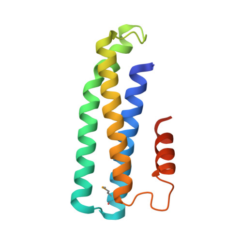
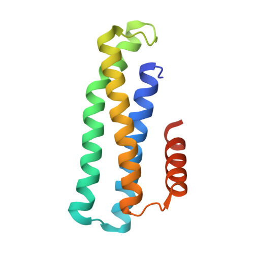
Crystal structure of PAV1-137: a protein from the virus PAV1 that infects Pyrococcus abyssi

Leulliot, N.Quevillon-Cheruel, S.Graille, M.Geslin, C.Flament, D.Romancer, M.L.Tilbeurgh, H.V.

(2013) Archaea 2013: 568053-568053

  • DOI: https://doi.org/10.1155/2013/568053
  • Primary Citation Related Structures: 
    4HR1

  • PubMed Abstract: 

    Pyrococcus abyssi virus 1 (PAV1) was the first virus particle infecting a hyperthermophilic Euryarchaeota (Pyrococcus abyssi strain GE23) that has been isolated and characterized. It is lemon shaped and is decorated with a short fibered tail. PAV1 morphologically resembles the fusiform members of the family Fuselloviridae or the genus Salterprovirus. The 18 kb dsDNA genome of PAV1 contains 25 predicted genes, most of them of unknown function. To help assigning functions to these proteins, we have initiated structural studies of the PAV1 proteome. We determined the crystal structure of a putative protein of 137 residues (PAV1-137) at a resolution of 2.2 Å. The protein forms dimers both in solution and in the crystal. The fold of PAV1-137 is a four- α -helical bundle analogous to those found in some eukaryotic adhesion proteins such as focal adhesion kinase, suggesting that PAV1-137 is involved in protein-protein interactions.


  • Organizational Affiliation
    • Institut de Biochimie et de Biophysique Moléculaire et Cellulaire, CNRS-UMR 8619, IFR115, Université Paris-Sud, Bâtiment 430, 91405 Orsay, France.

Macromolecule Content 

  • Total Structure Weight: 28.98 kDa 
  • Atom Count: 1,914 
  • Modeled Residue Count: 237 
  • Deposited Residue Count: 252 
  • Unique protein chains: 2

Macromolecules

Find similar proteins by:|  3D Structure
Entity ID: 1
MoleculeChains  Sequence LengthOrganismDetailsImage
Putative uncharacterized protein126Pyrococcus abyssi virus 1Mutation(s): 0 
Gene Names: ORF137Pav1-137
UniProt
Find proteins for A0ACD6B8E2 (Pyrococcus abyssi virus 1)
Explore A0ACD6B8E2 
Go to UniProtKB:  A0ACD6B8E2
Entity Groups
Sequence Clusters30% Identity50% Identity70% Identity90% Identity95% Identity100% Identity
UniProt GroupA0ACD6B8E2
Sequence Annotations
Expand
Reference Sequence
Find similar proteins by:|  3D Structure
Entity ID: 2
MoleculeChains  Sequence LengthOrganismDetailsImage
Putative uncharacterized protein126Pyrococcus abyssi virus 1Mutation(s): 0 
Gene Names: ORF137
UniProt
Find proteins for A0ACD6B8E2 (Pyrococcus abyssi virus 1)
Explore A0ACD6B8E2 
Go to UniProtKB:  A0ACD6B8E2
Entity Groups
Sequence Clusters30% Identity50% Identity70% Identity90% Identity95% Identity100% Identity
UniProt GroupA0ACD6B8E2
Sequence Annotations
Expand
Reference Sequence

Small Molecules

Modified Residues  1 Unique
IDChains TypeFormula2D DiagramParent
MSE
Query on MSE
A
L-PEPTIDE LINKINGC5 H11 N O2 SeMET

Experimental Data & Validation

Experimental Data

  • Method: X-RAY DIFFRACTION
  • Resolution: 2.50 Å
  • R-Value Free:  0.280 (Depositor), 0.277 (DCC) 
  • R-Value Work:  0.232 (Depositor) 
  • R-Value Observed: 0.234 (Depositor) 
Space Group: P 61 2 2
Unit Cell:
Length ( Å )Angle ( ˚ )
a = 73.5458α = 90
b = 73.5458β = 90
c = 191.5493γ = 120
Software Package:
Software NamePurpose
ADSCdata collection
SHARPphasing
BUSTERrefinement
MOSFLMdata reduction
SCALAdata scaling

Structure Validation

View Full Validation Report


Entry History 

Deposition Data

Revision History  (Full details and data files)

  • Version 1.0: 2013-06-19
    Type: Initial release
  • Version 1.1: 2024-11-13
    Changes: Data collection, Database references, Derived calculations, Structure summary