4HHW | pdb_00004hhw

Crystal structure of the Pseudomonas aeruginosa azurin, H124NO YOH122


Experimental Data Snapshot

  • Method: X-RAY DIFFRACTION
  • Resolution: 2.00 Å
  • R-Value Free: 
    0.281 (Depositor), 0.280 (DCC) 
  • R-Value Work: 
    0.224 (Depositor), 0.223 (DCC) 
  • R-Value Observed: 
    0.227 (Depositor) 

wwPDB Validation 3D Report Full Report

Validation slider image for 4HHW

This is version 1.3 of the entry. See complete history

Literature

Electron Flow through Nitrotyrosinate in Pseudomonas aeruginosa Azurin.

Warren, J.J.Herrera, N.Hill, M.G.Winkler, J.R.Gray, H.B.

(2013) J Am Chem Soc 135: 11151-11158

  • DOI: https://doi.org/10.1021/ja403734n
  • Primary Citation Related Structures: 
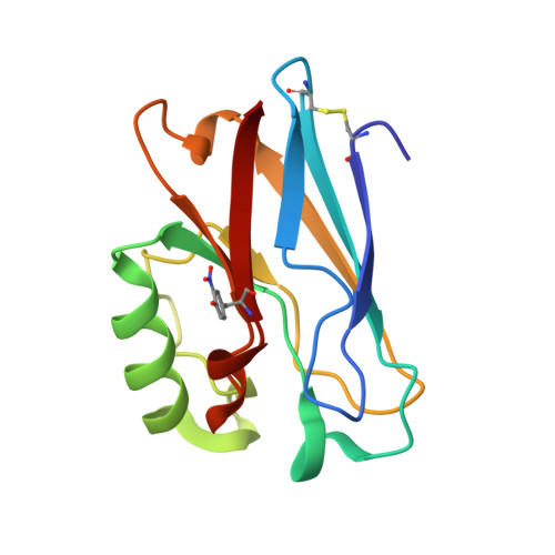
    4HHG, 4HHW, 4HIP

  • PubMed Abstract: 

    We have designed ruthenium-modified Pseudomonas aeruginosa azurins that incorporate 3-nitrotyrosine (NO2YOH) between Ru(2,2'-bipyridine)2(imidazole)(histidine) and Cu redox centers in electron transfer (ET) pathways. We investigated the structures and reactivities of three different systems: RuH107NO2YOH109, RuH124NO2YOH122, and RuH126NO2YOH122. RuH107NO2YOH109, unlabeled H124NO2YOH122, and unlabeled H126NO2YOH122 were structurally characterized. The pKa's of NO2YOH at positions 122 and 109 are 7.2 and 6.0, respectively. Reduction potentials of 3-nitrotyrosinate (NO2YO(-))-modified azurins were estimated from cyclic and differential pulse voltammetry data: oxidation of NO2YO(-)122 occurs near 1.1 versus NHE; oxidation of NO2YO(-)109 is near 1.2 V. Our analysis of transient optical spectroscopic experiments indicates that hopping via NO2YO(-) enhances Cu(I) oxidation rates over single-step ET by factors of 32 (RuH107NO2YO(-)109), 46 (RuH126NO2YO(-)122), and 13 (RuH124NO2YO(-)122).


  • Organizational Affiliation
    • Beckman Institute and Division of Chemistry and Chemical Engineering, California Institute of Technology, Pasadena, California 91125, USA.

Macromolecule Content 

  • Total Structure Weight: 28.12 kDa 
  • Atom Count: 2,007 
  • Modeled Residue Count: 256 
  • Deposited Residue Count: 256 
  • Unique protein chains: 1

Macromolecules

Find similar proteins by:|  3D Structure
Entity ID: 1
MoleculeChains  Sequence LengthOrganismDetailsImage
Azurin
A, B
128Pseudomonas aeruginosa PAO1Mutation(s): 6 
Gene Names: azuPA4922
UniProt
Find proteins for P00282 (Pseudomonas aeruginosa (strain ATCC 15692 / DSM 22644 / CIP 104116 / JCM 14847 / LMG 12228 / 1C / PRS 101 / PAO1))
Explore P00282 
Go to UniProtKB:  P00282
Entity Groups
Sequence Clusters30% Identity50% Identity70% Identity90% Identity95% Identity100% Identity
UniProt GroupP00282
Sequence Annotations
Expand
Reference Sequence

Small Molecules

Modified Residues  1 Unique
IDChains TypeFormula2D DiagramParent
NIY
Query on NIY
A, B
L-PEPTIDE LINKINGC9 H10 N2 O5TYR

Experimental Data & Validation

Experimental Data

  • Method: X-RAY DIFFRACTION
  • Resolution: 2.00 Å
  • R-Value Free:  0.281 (Depositor), 0.280 (DCC) 
  • R-Value Work:  0.224 (Depositor), 0.223 (DCC) 
  • R-Value Observed: 0.227 (Depositor) 
Space Group: P 2 21 21
Unit Cell:
Length ( Å )Angle ( ˚ )
a = 49.669α = 90
b = 65.637β = 90
c = 72.561γ = 90
Software Package:
Software NamePurpose
Blu-Icedata collection
MOLREPphasing
REFMACrefinement
autoXDSdata reduction
SCALAdata scaling

Structure Validation

View Full Validation Report


Entry History 

Deposition Data

Revision History  (Full details and data files)

  • Version 1.0: 2012-11-21
    Type: Initial release
  • Version 1.1: 2013-07-31
    Changes: Database references
  • Version 1.2: 2013-08-21
    Changes: Database references
  • Version 1.3: 2025-03-26
    Changes: Data collection, Database references, Derived calculations, Structure summary