4GAE | pdb_00004gae

Crystal structure of plasmodium dxr in complex with a pyridine-containing inhibitor


Experimental Data Snapshot

  • Method: X-RAY DIFFRACTION
  • Resolution: 2.30 Å
  • R-Value Free: 
    0.273 (Depositor), 0.273 (DCC) 
  • R-Value Work: 
    0.230 (Depositor), 0.230 (DCC) 
  • R-Value Observed: 
    0.230 (Depositor) 

Starting Model: experimental
View more details

wwPDB Validation 3D Report Full Report

Validation slider image for 4GAE

Ligand Structure Quality Assessment 


This is version 1.2 of the entry. See complete history

Literature

Antimalarial and Structural Studies of Pyridine-containing Inhibitors of 1-Deoxyxylulose-5-phosphate Reductoisomerase.

Xue, J.Diao, J.Cai, G.Deng, L.Zheng, B.Yao, Y.Song, Y.

(2013) ACS Med Chem Lett 4: 278-282

  • DOI: https://doi.org/10.1021/ml300419r
  • Primary Citation Related Structures: 
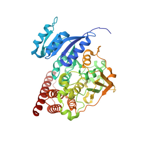
    4GAE

  • PubMed Abstract: 

    1-Deoxy- D -xylulose-5-phosphate reductoisomerase (DXR) in the non-mevalonate isoprene biosynthesis pathway is a target for developing antimalarial drugs. Fosmidomycin, a potent DXR inhibitor, showed safety as well as efficacy against P. falciparum malaria in clinical trials. Based on our previous quantitative structure activity relationship (QSAR) and crystallographic studies, several novel pyridine-containing fosmidomycin derivatives were designed, synthesized and found to be highly potent inhibitors of P. falciparum DXR ( Pf DXR) having K i values of 1.9 - 13 nM, with the best one being ~11× more active than fosmidomycin. These compounds also potently block the proliferation of multi-drug resistant P. falciparum with EC 50 values as low as 170 nM. A 2.3 Å crystal structure of Pf DXR in complex with one of the inhibitors is reported, showing the flexible loop of the protein undergoes conformational changes upon ligand binding and a hydrogen bond and favorable hydrophobic interactions between the pyridine group and Pf DXR account for the enhanced activity.


  • Organizational Affiliation
    • Department of Pharmacology, Baylor College of Medicine, 1 Baylor Plaza, Houston, Texas 77030, United States.

Macromolecule Content 

  • Total Structure Weight: 100.22 kDa 
  • Atom Count: 6,931 
  • Modeled Residue Count: 826 
  • Deposited Residue Count: 852 
  • Unique protein chains: 1

Macromolecules

Find similar proteins by:|  3D Structure
Entity ID: 1
MoleculeChains  Sequence LengthOrganismDetailsImage
1-deoxy-D-xylulose 5-phosphate reductoisomerase, apicoplast
A, B
426Plasmodium falciparum HB3Mutation(s): 1 
Gene Names: DXR
EC: 1.1.1.267
UniProt
Find proteins for O96693 (Plasmodium falciparum (isolate HB3))
Explore O96693 
Go to UniProtKB:  O96693
Entity Groups
Sequence Clusters30% Identity50% Identity70% Identity90% Identity95% Identity100% Identity
UniProt GroupO96693
Sequence Annotations
Expand
Reference Sequence

Small Molecules

Ligands 7 Unique
IDChains Name / Formula / InChI Key2D Diagram3D Interactions
NDP

Query on NDP


Download:Ideal Coordinates CCD File
C [auth A],
J [auth B]
NADPH DIHYDRO-NICOTINAMIDE-ADENINE-DINUCLEOTIDE PHOSPHATE
C21 H30 N7 O17 P3
ACFIXJIJDZMPPO-NNYOXOHSSA-N
FOQ

Query on FOQ


Download:Ideal Coordinates CCD File
E [auth A],
L [auth B]
[(1S)-3-[acetyl(hydroxy)amino]-1-(pyridin-4-yl)propyl]phosphonic acid
C10 H15 N2 O5 P
AMWIPWYKSYZXGB-JTQLQIEISA-N
FOB

Query on FOB


Download:Ideal Coordinates CCD File
D [auth A],
K [auth B]
[(1R)-3-[acetyl(hydroxy)amino]-1-(pyridin-4-yl)propyl]phosphonic acid
C10 H15 N2 O5 P
AMWIPWYKSYZXGB-SNVBAGLBSA-N
EDO

Query on EDO


Download:Ideal Coordinates CCD File
I [auth A],
Q [auth B],
R [auth B]
1,2-ETHANEDIOL
C2 H6 O2
LYCAIKOWRPUZTN-UHFFFAOYSA-N
ACT

Query on ACT


Download:Ideal Coordinates CCD File
H [auth A],
P [auth B]
ACETATE ION
C2 H3 O2
QTBSBXVTEAMEQO-UHFFFAOYSA-M
MN

Query on MN


Download:Ideal Coordinates CCD File
F [auth A],
M [auth B],
N [auth B],
O [auth B]
MANGANESE (II) ION
Mn
WAEMQWOKJMHJLA-UHFFFAOYSA-N
CL

Query on CL


Download:Ideal Coordinates CCD File
G [auth A]CHLORIDE ION
Cl
VEXZGXHMUGYJMC-UHFFFAOYSA-M

Experimental Data & Validation

Experimental Data

  • Method: X-RAY DIFFRACTION
  • Resolution: 2.30 Å
  • R-Value Free:  0.273 (Depositor), 0.273 (DCC) 
  • R-Value Work:  0.230 (Depositor), 0.230 (DCC) 
  • R-Value Observed: 0.230 (Depositor) 
Space Group: P 1 21 1
Unit Cell:
Length ( Å )Angle ( ˚ )
a = 51.36α = 90
b = 77.28β = 91.5
c = 109.41γ = 90
Software Package:
Software NamePurpose
StructureStudiodata collection
PHASERphasing
CNSrefinement
MOSFLMdata reduction
SCALAdata scaling

Structure Validation

View Full Validation Report


Ligand Structure Quality Assessment 


Entry History 

Deposition Data

Revision History  (Full details and data files)

  • Version 1.0: 2013-02-06
    Type: Initial release
  • Version 1.1: 2013-07-10
    Changes: Database references
  • Version 1.2: 2023-09-13
    Changes: Data collection, Database references, Derived calculations, Refinement description