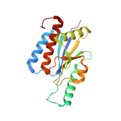
4FAK

Crystal Structure of OrfX in Complex with S-Adenosylmethionine


Experimental Data Snapshot

  • Method: X-RAY DIFFRACTION
  • Resolution: 1.70 Å
  • R-Value Free: 0.247 
  • R-Value Work: 0.207 
  • R-Value Observed: 0.207 

Starting Model: experimental
View more details

wwPDB Validation   3D Report Full Report


Ligand Structure Quality Assessment 


This is version 1.2 of the entry. See complete history


Literature

Characterization of the Staphylococcus aureus rRNA Methyltransferase Encoded by orfX, the Gene Containing the Staphylococcal Chromosome Cassette mec (SCCmec) Insertion Site.

Boundy, S.Safo, M.K.Wang, L.Musayev, F.N.O'Farrell, H.C.Rife, J.P.Archer, G.L.

(2013) J Biol Chem 288: 132-140

  • DOI: https://doi.org/10.1074/jbc.M112.385138
  • Primary Citation of Related Structures:  
    4FAK

  • PubMed Abstract: 

    The gene orfX is conserved among all staphylococci, and its complete sequence is maintained upon insertion of the staphylococcal chromosome cassette mec (SCCmec) genomic island, containing the gene encoding resistance to β-lactam antibiotics (mecA), into its C terminus. The function of OrfX has not been determined. We show that OrfX was constitutively produced during growth, that orfX could be inactivated without altering bacterial growth, and that insertion of SCCmec did not alter gene expression. We solved the crystal structure of OrfX at 1.7 Å and found that it belongs to the S-adenosyl-L-methionine (AdoMet)-dependent α/β-knot superfamily of SPOUT methyltransferases (MTases), with a high structural homology to YbeA, the gene product of the Escherichia coli 70 S ribosomal MTase RlmH. MTase activity was confirmed by demonstrating the OrfX-dependent methylation of the Staphylococcus aureus 70 S ribosome. When OrfX was crystallized in the presence of its AdoMet substrate, we found that each monomer of the homodimeric structure bound AdoMet in its active site. Solution studies using isothermal titration calorimetry confirmed that each monomer bound AdoMet but with different binding affinities (K(d) = 52 ± 0.4 and 606 ± 2 μm). In addition, the structure shows that the AdoMet-binding pocket, formed by a deep trefoil knot, contains a bound phosphate molecule, which is the likely nucleotide methylation site. This study represents the first characterization of a staphylococcal ribosomal MTase and provides the first crystal structure of a member of the α/β-knot superfamily of SPOUT MTases in the RlmH or COG1576 family with bound AdoMet.


  • Organizational Affiliation

    Division of Infectious Diseases, Department of Medicine, School of Medicine, Virginia Commonwealth University, Richmond, Virginia 23298 USA.


Macromolecules
Find similar proteins by:  (by identity cutoff)  |  3D Structure
Entity ID: 1
MoleculeChains Sequence LengthOrganismDetailsImage
Ribosomal RNA large subunit methyltransferase H163Staphylococcus aureusMutation(s): 0 
Gene Names: orfxrlmH
EC: 2.1.1.177
UniProt
Find proteins for P0C1V0 (Staphylococcus aureus)
Explore P0C1V0 
Go to UniProtKB:  P0C1V0
Entity Groups  
Sequence Clusters30% Identity50% Identity70% Identity90% Identity95% Identity100% Identity
UniProt GroupP0C1V0
Sequence Annotations
Expand
  • Reference Sequence
Small Molecules
Ligands 3 Unique
IDChains Name / Formula / InChI Key2D Diagram3D Interactions
SAM
Query on SAM

Download Ideal Coordinates CCD File 
B [auth A]S-ADENOSYLMETHIONINE
C15 H22 N6 O5 S
MEFKEPWMEQBLKI-FCKMPRQPSA-N
PG4
Query on PG4

Download Ideal Coordinates CCD File 
C [auth A]TETRAETHYLENE GLYCOL
C8 H18 O5
UWHCKJMYHZGTIT-UHFFFAOYSA-N
PO4
Query on PO4

Download Ideal Coordinates CCD File 
D [auth A]PHOSPHATE ION
O4 P
NBIIXXVUZAFLBC-UHFFFAOYSA-K
Binding Affinity Annotations 
IDSourceBinding Affinity
SAM PDBBind:  4FAK Kd: 5.20e+4 (nM) from 1 assay(s)
Experimental Data & Validation

Experimental Data

  • Method: X-RAY DIFFRACTION
  • Resolution: 1.70 Å
  • R-Value Free: 0.247 
  • R-Value Work: 0.207 
  • R-Value Observed: 0.207 
  • Space Group: C 2 2 21
Unit Cell:
Length ( Å )Angle ( ˚ )
a = 77.8α = 90
b = 84.29β = 90
c = 55.73γ = 90
Software Package:
Software NamePurpose
CrystalCleardata collection
PHASERphasing
CNSrefinement
CrystalCleardata reduction
CrystalCleardata scaling

Structure Validation

View Full Validation Report


Ligand Structure Quality Assessment 


Entry History 

Deposition Data

Revision History  (Full details and data files)

  • Version 1.0: 2012-06-06
    Type: Initial release
  • Version 1.1: 2013-01-30
    Changes: Database references
  • Version 1.2: 2023-09-13
    Changes: Data collection, Database references, Derived calculations, Refinement description