4ESV | pdb_00004esv

A New Twist on the Translocation Mechanism of Helicases from the Structure of DnaB with its Substrates


Experimental Data Snapshot

  • Method: X-RAY DIFFRACTION
  • Resolution: 3.20 Å
  • R-Value Free: 
    0.289 (Depositor), 0.281 (DCC) 
  • R-Value Work: 
    0.242 (Depositor), 0.239 (DCC) 
  • R-Value Observed: 
    0.245 (Depositor) 

wwPDB Validation 3D Report Full Report

Validation slider image for 4ESV

Ligand Structure Quality Assessment 


This is version 1.3 of the entry. See complete history

Literature

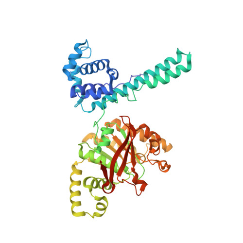
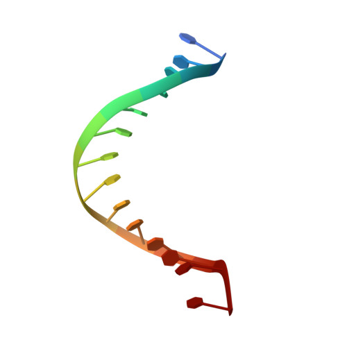
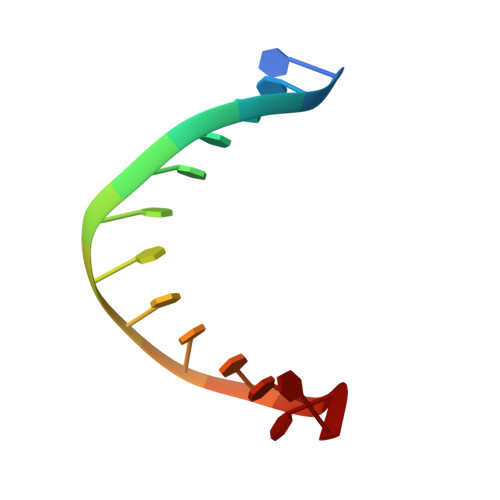
The Hexameric Helicase DnaB Adopts a Nonplanar Conformation during Translocation.

Itsathitphaisarn, O.Wing, R.A.Eliason, W.K.Wang, J.Steitz, T.A.

(2012) Cell 151: 267-277

  • DOI: https://doi.org/10.1016/j.cell.2012.09.014
  • Primary Citation Related Structures: 
    4ESV

  • PubMed Abstract: 

    DNA polymerases can only synthesize nascent DNA from single-stranded DNA (ssDNA) templates. In bacteria, the unwinding of parental duplex DNA is carried out by the replicative DNA helicase (DnaB) that couples NTP hydrolysis to 5' to 3' translocation. The crystal structure of the DnaB hexamer in complex with GDP-AlF(4) and ssDNA reported here reveals that DnaB adopts a closed spiral staircase quaternary structure around an A-form ssDNA with each C-terminal domain coordinating two nucleotides of ssDNA. The structure not only provides structural insights into the translocation mechanism of superfamily IV helicases but also suggests that members of this superfamily employ a translocation mechanism that is distinct from other helicase superfamilies. We propose a hand-over-hand mechanism in which sequential hydrolysis of NTP causes a sequential 5' to 3' movement of the subunits along the helical axis of the staircase, resulting in the unwinding of two nucleotides per subunit.


  • Organizational Affiliation
    • Department of Molecular Biophysics and Biochemistry, Yale University, New Haven, CT 06520, USA.

Macromolecule Content 

  • Total Structure Weight: 623.32 kDa 
  • Atom Count: 40,296 
  • Modeled Residue Count: 5,099 
  • Deposited Residue Count: 5,475 
  • Unique protein chains: 1
  • Unique nucleic acid chains: 2

Macromolecules


Find similar proteins by:|  3D Structure
Entity ID: 3
MoleculeChains  Sequence LengthOrganismDetailsImage
Replicative helicase454Geobacillus stearothermophilusMutation(s): 0 
Gene Names: dnaB
EC: 3.6.4.12 (PDB Primary Data), 5.6.2.3 (UniProt)
UniProt
Find proteins for Q9X4C9 (Geobacillus stearothermophilus)
Explore Q9X4C9 
Go to UniProtKB:  Q9X4C9
Entity Groups
Sequence Clusters30% Identity50% Identity70% Identity90% Identity95% Identity100% Identity
UniProt GroupQ9X4C9
Sequence Annotations
Expand
Reference Sequence
Find similar nucleic acids by:  Sequence
Entity ID: 1
MoleculeChains LengthOrganismImage
5'-D(P*TP*TP*TP*TP*TP*TP*TP*TP*TP*TP*TP*TP*TP*T)-3'A [auth V]14synthetic construct
Sequence Annotations
Expand
Reference Sequence
Find similar nucleic acids by:  Sequence
Entity ID: 2
MoleculeChains LengthOrganismImage
5'-D(P*TP*TP*TP*TP*TP*TP*TP*TP*TP*TP*TP*TP*T)-3'B [auth W]13synthetic construct
Sequence Annotations
Expand
Reference Sequence

Small Molecules

Ligands 4 Unique
IDChains Name / Formula / InChI Key2D Diagram3D Interactions
GDP

Query on GDP


Download:Ideal Coordinates CCD File
CA [auth D]
GA [auth E]
KA [auth F]
MA [auth G]
PA [auth I]
CA [auth D],
GA [auth E],
KA [auth F],
MA [auth G],
PA [auth I],
Q [auth A],
TA [auth J],
VA [auth K],
X [auth C],
XA [auth L]
GUANOSINE-5'-DIPHOSPHATE
C10 H15 N5 O11 P2
QGWNDRXFNXRZMB-UUOKFMHZSA-N
MES

Query on MES


Download:Ideal Coordinates CCD File
BA [auth C],
JA [auth E],
O [auth W],
SA [auth I]
2-(N-MORPHOLINO)-ETHANESULFONIC ACID
C6 H13 N O4 S
SXGZJKUKBWWHRA-UHFFFAOYSA-N
ALF

Query on ALF


Download:Ideal Coordinates CCD File
AA [auth C]
EA [auth D]
IA [auth E]
NA [auth G]
RA [auth I]
AA [auth C],
EA [auth D],
IA [auth E],
NA [auth G],
RA [auth I],
S [auth A],
UA [auth J],
WA [auth K],
YA [auth L],
Z [auth C]
TETRAFLUOROALUMINATE ION
Al F4
UYOMQIYKOOHAMK-UHFFFAOYSA-J
CA

Query on CA


Download:Ideal Coordinates CCD File
DA [auth D]
FA [auth D]
HA [auth E]
LA [auth F]
OA [auth G]
DA [auth D],
FA [auth D],
HA [auth E],
LA [auth F],
OA [auth G],
P [auth A],
QA [auth I],
R [auth A],
T [auth A],
U [auth A],
V [auth A],
W [auth B],
Y [auth C],
ZA [auth L]
CALCIUM ION
Ca
BHPQYMZQTOCNFJ-UHFFFAOYSA-N

Experimental Data & Validation

Experimental Data

  • Method: X-RAY DIFFRACTION
  • Resolution: 3.20 Å
  • R-Value Free:  0.289 (Depositor), 0.281 (DCC) 
  • R-Value Work:  0.242 (Depositor), 0.239 (DCC) 
  • R-Value Observed: 0.245 (Depositor) 
Space Group: P 2 2 21
Unit Cell:
Length ( Å )Angle ( ˚ )
a = 149.124α = 90
b = 180.321β = 90
c = 279.128γ = 90
Software Package:
Software NamePurpose
DENZOdata reduction
SCALEPACKdata scaling
SHELXphasing
SHELXmodel building
REFMACrefinement
PDB_EXTRACTdata extraction
ADSCdata collection
CBASSdata collection
HKL-2000data reduction
HKL-2000data scaling
SHELXDphasing

Structure Validation

View Full Validation Report


Ligand Structure Quality Assessment 


Entry History 

Deposition Data

Revision History  (Full details and data files)

  • Version 1.0: 2012-10-24
    Type: Initial release
  • Version 1.1: 2017-11-15
    Changes: Advisory, Refinement description
  • Version 1.2: 2018-06-06
    Changes: Data collection, Refinement description, Source and taxonomy
  • Version 1.3: 2024-02-28
    Changes: Advisory, Data collection, Database references, Derived calculations