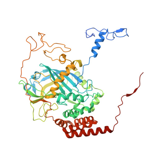
4CAB

The refined structure of catalase DR1998 from Deinococcus radiodurans at 2.6 A resolution


Experimental Data Snapshot

  • Method: X-RAY DIFFRACTION
  • Resolution: 2.60 Å
  • R-Value Free: 0.236 
  • R-Value Work: 0.236 
  • R-Value Observed: 0.236 

Starting Models: experimental
View more details

wwPDB Validation   3D Report Full Report


Ligand Structure Quality Assessment 


This is version 1.3 of the entry. See complete history


Literature

Structure of the Mono-Functional Heme Catalase Dr1998 from Deinococcus Radiodurans

Borges, P.T.Frazao, C.Miranda, C.S.Carrondo, M.A.Romao, C.V.

(2014) FEBS J 281: 4138

  • DOI: https://doi.org/10.1111/febs.12895
  • Primary Citation of Related Structures:  
    4CAB

  • PubMed Abstract: 

    Deinococcus radiodurans is an aerobic organism with the ability to survive under conditions of high radiation doses or desiccation. As part of its protection system against oxidative stress, this bacterium encodes three monofunctional catalases. The DR1998 catalase belongs to clade 1, and is present at high levels under normal growth conditions. The crystals of DR1998 diffracted very weakly, and the merged diffraction data showed an R sym of 0.308. Its crystal structure was determined and refined to 2.6 Å. The four molecules present in the asymmetric unit form, by crystallographic symmetry, two homotetramers with 222 point-group symmetry. The overall structure of DR1998 is similar to that of other monofunctional catalases, showing higher structural homology with the catalase structures of clade 1. Each monomer shows the typical catalase fold, and contains one heme b in the active site. The heme is coordinated by the proximal ligand Tyr369, and on the heme distal side the essential His81 and Asn159 are hydrogen-bonded to a water molecule. A 25-Å-long channel is the main channel connecting the active site to the external surface. This channel starts with a hydrophobic region from the catalytic heme site, which is followed by a hydrophilic region that begins on Asp139 and expands up to the protein surface. Apart from this channel, an alternative channel, also near the heme active site, is presented and discussed. Coordinates and structure factors have been deposited in the Protein Data Bank in Europe under accession code 4CAB.


  • Organizational Affiliation

    Instituto de Tecnologia Química e Biológica, Universidade Nova de Lisboa, Oeiras, Portugal.


Macromolecules
Find similar proteins by:  (by identity cutoff)  |  3D Structure
Entity ID: 1
MoleculeChains Sequence LengthOrganismDetailsImage
CATALASE
A, B, C, D
536Deinococcus radiodurans R1 = ATCC 13939 = DSM 20539Mutation(s): 0 
EC: 1.11.1.6
UniProt
Find proteins for Q59337 (Deinococcus radiodurans (strain ATCC 13939 / DSM 20539 / JCM 16871 / CCUG 27074 / LMG 4051 / NBRC 15346 / NCIMB 9279 / VKM B-1422 / R1))
Explore Q59337 
Go to UniProtKB:  Q59337
Entity Groups  
Sequence Clusters30% Identity50% Identity70% Identity90% Identity95% Identity100% Identity
UniProt GroupQ59337
Sequence Annotations
Expand
  • Reference Sequence
Small Molecules
Ligands 2 Unique
IDChains Name / Formula / InChI Key2D Diagram3D Interactions
HEM
Query on HEM

Download Ideal Coordinates CCD File 
E [auth A],
I [auth B],
M [auth C],
Q [auth D]
PROTOPORPHYRIN IX CONTAINING FE
C34 H32 Fe N4 O4
KABFMIBPWCXCRK-RGGAHWMASA-L
CL
Query on CL

Download Ideal Coordinates CCD File 
F [auth A]
G [auth A]
H [auth A]
J [auth B]
K [auth B]
F [auth A],
G [auth A],
H [auth A],
J [auth B],
K [auth B],
L [auth B],
N [auth C],
O [auth C],
P [auth C],
R [auth D]
CHLORIDE ION
Cl
VEXZGXHMUGYJMC-UHFFFAOYSA-M
Experimental Data & Validation

Experimental Data

  • Method: X-RAY DIFFRACTION
  • Resolution: 2.60 Å
  • R-Value Free: 0.236 
  • R-Value Work: 0.236 
  • R-Value Observed: 0.236 
  • Space Group: C 2 2 21
Unit Cell:
Length ( Å )Angle ( ˚ )
a = 97.326α = 90
b = 311.875β = 90
c = 145.628γ = 90
Software Package:
Software NamePurpose
PHENIXrefinement
XDSdata reduction
PHASERphasing

Structure Validation

View Full Validation Report


Ligand Structure Quality Assessment 


Entry History 

Deposition Data

Revision History  (Full details and data files)

  • Version 1.0: 2014-07-09
    Type: Initial release
  • Version 1.1: 2015-08-12
    Changes: Database references
  • Version 1.2: 2019-02-27
    Changes: Data collection, Experimental preparation, Other
  • Version 1.3: 2023-12-20
    Changes: Data collection, Database references, Derived calculations, Other, Refinement description