4CA1

Crystal structure of Siah1 at 1.58 A resolution.


Experimental Data Snapshot

  • Method: X-RAY DIFFRACTION
  • Resolution: 1.58 Å
  • R-Value Free: 0.189 
  • R-Value Work: 0.141 
  • R-Value Observed: 0.143 

wwPDB Validation   3D Report Full Report


This is version 1.3 of the entry. See complete history


Literature

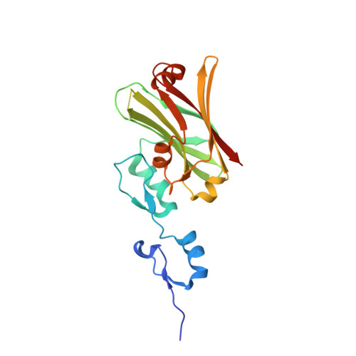
Two High-Resolution Structures of the Human E3 Ubiquitin Ligase Siah1.

Rimsa, V.Eadsforth, T.C.Hunter, W.N.

(2013) Acta Crystallogr Sect F Struct Biol Cryst Commun 96: 1339

  • DOI: https://doi.org/10.1107/S1744309113031448
  • Primary Citation of Related Structures:  
    4C9Z, 4CA1

  • PubMed Abstract: 

    Siah1 is an E3 ubiquitin ligase that contributes to proteasome-mediated degradation of multiple targets in key cellular processes and which shows promise as a therapeutic target in oncology. Structures of a truncated Siah1 bound to peptide-based inhibitors have been reported. Here, new crystallization conditions have allowed the determination of a construct encompassing dual zinc-finger subdomains and substrate-binding domains at significantly higher resolution. Although the crystals appear isomorphous, two structures present distinct states in which the spatial orientation of one zinc-finger subdomain differs with respect to the rest of the dimeric protein. Such a difference, which is indicative of conformational freedom, infers potential biological relevance related to recognition of binding partners. The crystallization conditions and improved models of Siah1 may aid future studies investigating Siah1-ligand complexes.


  • Organizational Affiliation

    Division of Biological Chemistry and Drug Discovery, College of Life Sciences, University of Dundee, Dundee DD1 5EH, Scotland.


Macromolecules
Find similar proteins by:  (by identity cutoff)  |  3D Structure
Entity ID: 1
MoleculeChains Sequence LengthOrganismDetailsImage
E3 UBIQUITIN-PROTEIN LIGASE SIAH1
A, B
195Homo sapiensMutation(s): 0 
EC: 6.3.2
UniProt & NIH Common Fund Data Resources
Find proteins for Q8IUQ4 (Homo sapiens)
Explore Q8IUQ4 
Go to UniProtKB:  Q8IUQ4
PHAROS:  Q8IUQ4
GTEx:  ENSG00000196470 
Entity Groups  
Sequence Clusters30% Identity50% Identity70% Identity90% Identity95% Identity100% Identity
UniProt GroupQ8IUQ4
Sequence Annotations
Expand
  • Reference Sequence
Small Molecules
Ligands 4 Unique
IDChains Name / Formula / InChI Key2D Diagram3D Interactions
SO4
Query on SO4

Download Ideal Coordinates CCD File 
E [auth A],
F [auth A],
G [auth A],
R [auth B],
S [auth B]
SULFATE ION
O4 S
QAOWNCQODCNURD-UHFFFAOYSA-L
GOL
Query on GOL

Download Ideal Coordinates CCD File 
I [auth A]
J [auth A]
K [auth A]
L [auth A]
M [auth A]
I [auth A],
J [auth A],
K [auth A],
L [auth A],
M [auth A],
N [auth A],
U [auth B],
V [auth B],
W [auth B],
X [auth B],
Y [auth B],
Z [auth B]
GLYCEROL
C3 H8 O3
PEDCQBHIVMGVHV-UHFFFAOYSA-N
ZN
Query on ZN

Download Ideal Coordinates CCD File 
C [auth A],
D [auth A],
P [auth B],
Q [auth B]
ZINC ION
Zn
PTFCDOFLOPIGGS-UHFFFAOYSA-N
CL
Query on CL

Download Ideal Coordinates CCD File 
H [auth A],
O [auth A],
T [auth B]
CHLORIDE ION
Cl
VEXZGXHMUGYJMC-UHFFFAOYSA-M
Experimental Data & Validation

Experimental Data

  • Method: X-RAY DIFFRACTION
  • Resolution: 1.58 Å
  • R-Value Free: 0.189 
  • R-Value Work: 0.141 
  • R-Value Observed: 0.143 
  • Space Group: I 2 2 2
Unit Cell:
Length ( Å )Angle ( ˚ )
a = 76.06α = 90
b = 104.12β = 90
c = 133.59γ = 90
Software Package:
Software NamePurpose
REFMACrefinement
iMOSFLMdata reduction
Aimlessdata scaling
PHASERphasing

Structure Validation

View Full Validation Report


Entry History 

Deposition Data

Revision History  (Full details and data files)

  • Version 1.0: 2013-10-16
    Type: Initial release
  • Version 1.1: 2013-12-11
    Changes: Database references
  • Version 1.2: 2013-12-18
    Changes: Database references
  • Version 1.3: 2023-12-20
    Changes: Data collection, Database references, Derived calculations, Other, Refinement description