4C65 | pdb_00004c65

Crystal structure of A. niger ochratoxinase


Experimental Data Snapshot

  • Method: X-RAY DIFFRACTION
  • Resolution: 2.20 Å
  • R-Value Free: 
    0.245 (Depositor), 0.250 (DCC) 
  • R-Value Work: 
    0.213 (Depositor), 0.217 (DCC) 
  • R-Value Observed: 
    0.215 (Depositor) 

Starting Model: experimental
View more details

wwPDB Validation 3D Report Full Report

Validation slider image for 4C65

This is version 1.2 of the entry. See complete history

Literature

Structural and Functional Characterization of Ochratoxinase, a Novel Mycotoxin Degrading Enzyme.

Dobritzsch, D.Wang, H.Schneider, G.Yu, S.

(2014) Biochem J 462: 441

  • DOI: https://doi.org/10.1042/BJ20140382
  • Primary Citation Related Structures: 
    4C5Y, 4C5Z, 4C60, 4C65

  • PubMed Abstract: 

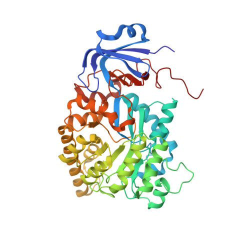
    Ochratoxin, with ochratoxin A as the dominant form, is one of the five major mycotoxins most harmful to humans and animals. It is produced by Aspergillus and Penicillium species and occurs in a wide range of agricultural products. Detoxification of contaminated food is a challenging health issue. In the present paper we report the identification, characterization and crystal structure (at 2.2 Å) of a novel microbial ochratoxinase from Aspergillus niger. A putative amidase gene encoding a 480 amino acid polypeptide was cloned and homologously expressed in A. niger. The recombinant protein is N-terminally truncated, thermostable, has optimal activity at pH ~6 and 66°C, and is more efficient in ochratoxin A hydrolysis than carboxypeptidase A and Y, the two previously known enzymes capable of degrading this mycotoxin. The subunit of the homo-octameric enzyme folds into a two-domain structure characteristic of a metal dependent amidohydrolase, with a twisted TIM (triosephosphateisomerase)-barrel and a smaller β-sandwich domain. The active site contains an aspartate residue for acid-base catalysis, and a carboxylated lysine and four histidine residues for binding of a binuclear metal centre.


  • Organizational Affiliation
    • *Department of Medical Biochemistry and Biophysics, Karolinska Institutet, SE-17177 Stockholm, Sweden.

Macromolecule Content 

  • Total Structure Weight: 373.17 kDa 
  • Atom Count: 27,300 
  • Modeled Residue Count: 3,463 
  • Deposited Residue Count: 3,504 
  • Unique protein chains: 1

Macromolecules

Find similar proteins by:|  3D Structure
Entity ID: 1
MoleculeChains  Sequence LengthOrganismDetailsImage
OCHRATOXINASE
A, B, C, D, E
A, B, C, D, E, F, G, H
438Aspergillus nigerMutation(s): 0 
EC: 3.4.13.9 (PDB Primary Data), 3.5.1 (PDB Primary Data), 3.4.17 (UniProt)
UniProt
Find proteins for A2R2V4 (Aspergillus niger (strain ATCC MYA-4892 / CBS 513.88 / FGSC A1513))
Explore A2R2V4 
Go to UniProtKB:  A2R2V4
Entity Groups
Sequence Clusters30% Identity50% Identity70% Identity90% Identity95% Identity100% Identity
UniProt GroupA2R2V4
Sequence Annotations
Expand
Reference Sequence

Experimental Data & Validation

Experimental Data

  • Method: X-RAY DIFFRACTION
  • Resolution: 2.20 Å
  • R-Value Free:  0.245 (Depositor), 0.250 (DCC) 
  • R-Value Work:  0.213 (Depositor), 0.217 (DCC) 
  • R-Value Observed: 0.215 (Depositor) 
Space Group: I 1 2 1
Unit Cell:
Length ( Å )Angle ( ˚ )
a = 210.855α = 90
b = 78.92β = 105.05
c = 217γ = 90
Software Package:
Software NamePurpose
REFMACrefinement
iMOSFLMdata reduction
SCALAdata scaling
REFMACphasing

Structure Validation

View Full Validation Report


Entry History 

Deposition Data

Revision History  (Full details and data files)

  • Version 1.0: 2014-07-02
    Type: Initial release
  • Version 1.1: 2014-09-03
    Changes: Database references
  • Version 1.2: 2023-12-20
    Changes: Data collection, Database references, Other, Refinement description