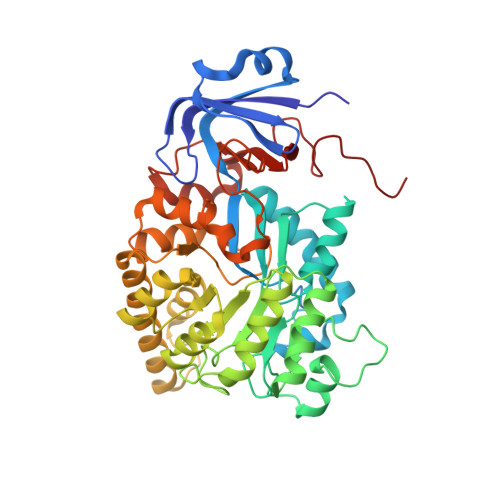
4C65

Crystal structure of A. niger ochratoxinase


Experimental Data Snapshot

  • Method: X-RAY DIFFRACTION
  • Resolution: 2.20 Å
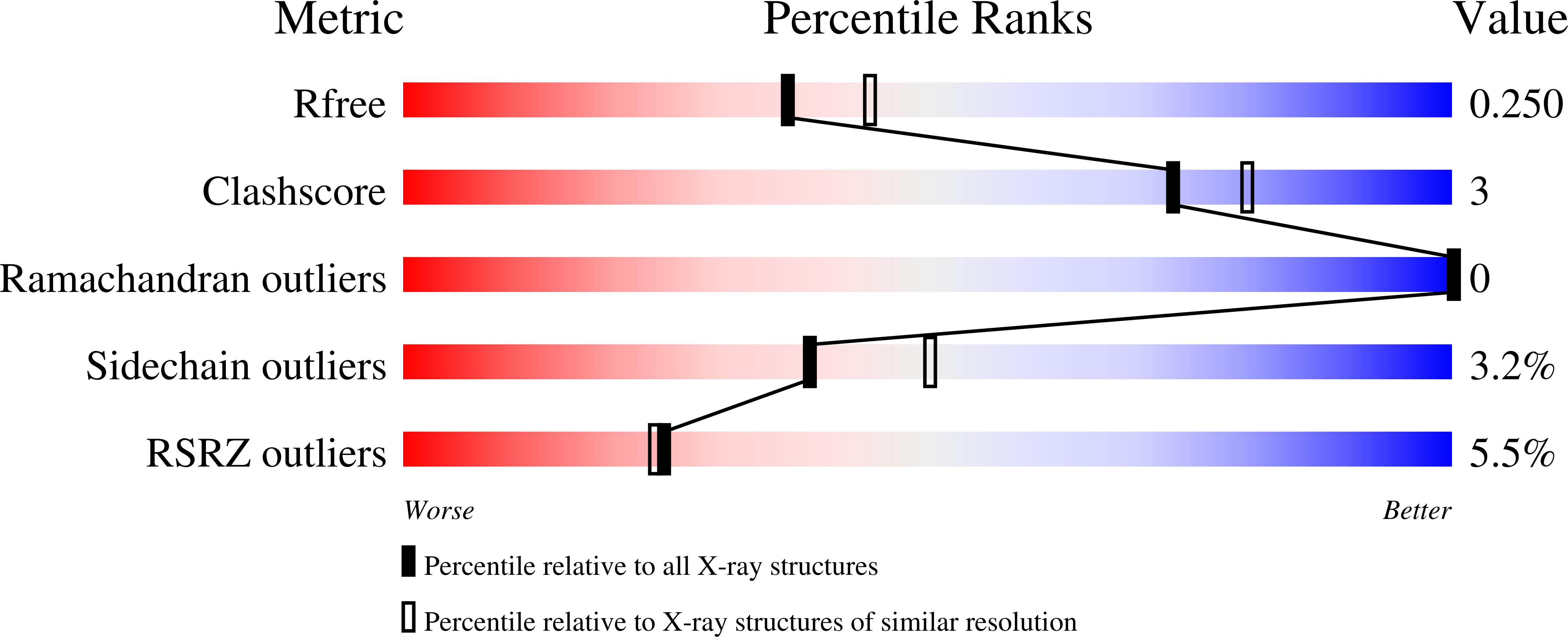
  • R-Value Free: 0.245 
  • R-Value Work: 0.213 
  • R-Value Observed: 0.215 

Starting Model: experimental
View more details

wwPDB Validation   3D Report Full Report


This is version 1.2 of the entry. See complete history


Literature

Structural and Functional Characterization of Ochratoxinase, a Novel Mycotoxin Degrading Enzyme.

Dobritzsch, D.Wang, H.Schneider, G.Yu, S.

(2014) Biochem J 462: 441

  • DOI: https://doi.org/10.1042/BJ20140382
  • Primary Citation of Related Structures:  
    4C5Y, 4C5Z, 4C60, 4C65

  • PubMed Abstract: 

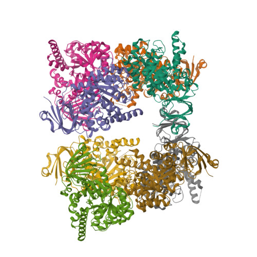
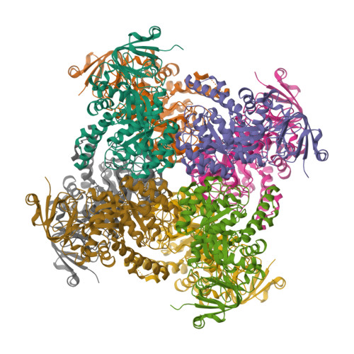
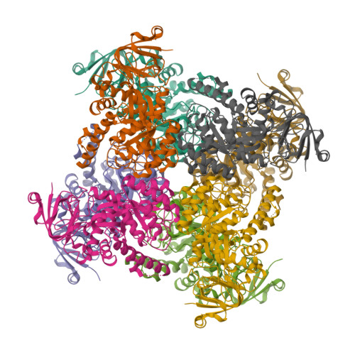
    Ochratoxin, with ochratoxin A as the dominant form, is one of the five major mycotoxins most harmful to humans and animals. It is produced by Aspergillus and Penicillium species and occurs in a wide range of agricultural products. Detoxification of contaminated food is a challenging health issue. In the present paper we report the identification, characterization and crystal structure (at 2.2 Å) of a novel microbial ochratoxinase from Aspergillus niger. A putative amidase gene encoding a 480 amino acid polypeptide was cloned and homologously expressed in A. niger. The recombinant protein is N-terminally truncated, thermostable, has optimal activity at pH ~6 and 66°C, and is more efficient in ochratoxin A hydrolysis than carboxypeptidase A and Y, the two previously known enzymes capable of degrading this mycotoxin. The subunit of the homo-octameric enzyme folds into a two-domain structure characteristic of a metal dependent amidohydrolase, with a twisted TIM (triosephosphateisomerase)-barrel and a smaller β-sandwich domain. The active site contains an aspartate residue for acid-base catalysis, and a carboxylated lysine and four histidine residues for binding of a binuclear metal centre.


  • Organizational Affiliation

    *Department of Medical Biochemistry and Biophysics, Karolinska Institutet, SE-17177 Stockholm, Sweden.


Macromolecules
Find similar proteins by:  (by identity cutoff)  |  3D Structure
Entity ID: 1
MoleculeChains Sequence LengthOrganismDetailsImage
OCHRATOXINASE
A, B, C, D, E
A, B, C, D, E, F, G, H
438Aspergillus nigerMutation(s): 0 
EC: 3.4.13.9 (PDB Primary Data), 3.5.1 (PDB Primary Data), 3.4.17 (UniProt)
UniProt
Find proteins for A2R2V4 (Aspergillus niger (strain ATCC MYA-4892 / CBS 513.88 / FGSC A1513))
Explore A2R2V4 
Go to UniProtKB:  A2R2V4
Entity Groups  
Sequence Clusters30% Identity50% Identity70% Identity90% Identity95% Identity100% Identity
UniProt GroupA2R2V4
Sequence Annotations
Expand
  • Reference Sequence
Experimental Data & Validation

Experimental Data

  • Method: X-RAY DIFFRACTION
  • Resolution: 2.20 Å
  • R-Value Free: 0.245 
  • R-Value Work: 0.213 
  • R-Value Observed: 0.215 
  • Space Group: I 1 2 1
Unit Cell:
Length ( Å )Angle ( ˚ )
a = 210.855α = 90
b = 78.92β = 105.05
c = 217γ = 90
Software Package:
Software NamePurpose
REFMACrefinement
iMOSFLMdata reduction
SCALAdata scaling
REFMACphasing

Structure Validation

View Full Validation Report


Entry History 

Deposition Data

Revision History  (Full details and data files)

  • Version 1.0: 2014-07-02
    Type: Initial release
  • Version 1.1: 2014-09-03
    Changes: Database references
  • Version 1.2: 2023-12-20
    Changes: Data collection, Database references, Other, Refinement description