4BP3 | pdb_00004bp3

Crystal Structure of Plasmodium Falciparum Spermidine Synthase in Complex with DECARBOXYLATED S-ADENOSYLMETHIONINE5' AND 4- METHYLANILINE


Experimental Data Snapshot

  • Method: X-RAY DIFFRACTION
  • Resolution: 1.75 Å
  • R-Value Free: 
    0.212 (Depositor), 0.218 (DCC) 
  • R-Value Work: 
    0.180 (Depositor), 0.187 (DCC) 
  • R-Value Observed: 
    0.181 (Depositor) 

Starting Model: experimental
View more details

wwPDB Validation 3D Report Full Report

Validation slider image for 4BP3

Ligand Structure Quality Assessment 


This is version 1.5 of the entry. See complete history

Literature

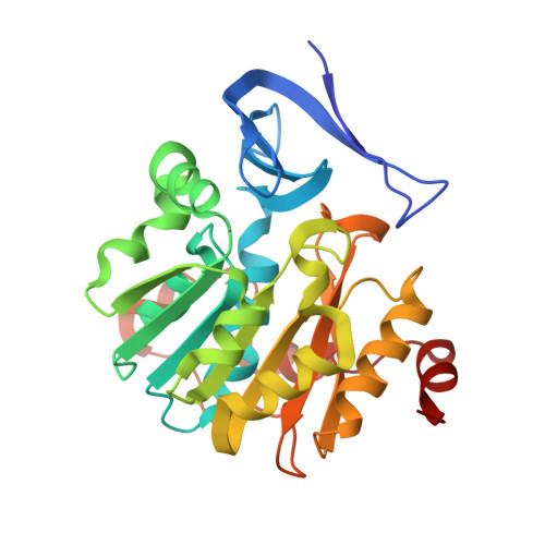
Three-Dimensional Structures of Plasmodium Falciparum Spermidine Synthase with Bound Inhibitors Suggest New Strategies for Drug Design

Sprenger, J.Svensson, B.Halander, J.Carey, J.Persson, L.Al-Karadaghi, S.

(2015) Acta Crystallogr D Biol Crystallogr 71: 484

  • DOI: https://doi.org/10.1107/S1399004714027011
  • Primary Citation Related Structures: 
    4BP1, 4BP3, 4CWA, 4CXM, 4UOE

  • PubMed Abstract: 

    The enzymes of the polyamine-biosynthesis pathway have been proposed to be promising drug targets in the treatment of malaria. Spermidine synthase (SpdS; putrescine aminopropyltransferase) catalyzes the transfer of the aminopropyl moiety from decarboxylated S-adenosylmethionine to putrescine, leading to the formation of spermidine and 5'-methylthioadenosine (MTA). In this work, X-ray crystallography was used to examine ligand complexes of SpdS from the malaria parasite Plasmodium falciparum (PfSpdS). Five crystal structures were determined of PfSpdS in complex with MTA and the substrate putrescine, with MTA and spermidine, which was obtained as a result of the enzymatic reaction taking place within the crystals, with dcAdoMet and the inhibitor 4-methylaniline, with MTA and 4-aminomethylaniline, and with a compound predicted in earlier in silico screening to bind to the active site of the enzyme, benzimidazol-(2-yl)pentan-1-amine (BIPA). In contrast to the other inhibitors tested, the complex with BIPA was obtained without any ligand bound to the dcAdoMet-binding site of the enzyme. The complexes with the aniline compounds and BIPA revealed a new mode of ligand binding to PfSpdS. The observed binding mode of the ligands, and the interplay between the two substrate-binding sites and the flexible gatekeeper loop, can be used in the design of new approaches in the search for new inhibitors of SpdS.


  • Organizational Affiliation
    • Center for Molecular Protein Science, Lund University, SE-221 00 Lund, Sweden.

Macromolecule Content 

  • Total Structure Weight: 98.65 kDa 
  • Atom Count: 7,288 
  • Modeled Residue Count: 842 
  • Deposited Residue Count: 849 
  • Unique protein chains: 1

Macromolecules

Find similar proteins by:|  3D Structure
Entity ID: 1
MoleculeChains  Sequence LengthOrganismDetailsImage
SPERMIDINE SYNTHASE
A, B, C
283Plasmodium falciparum 3D7Mutation(s): 0 
EC: 2.5.1.16
UniProt
Find proteins for Q8II73 (Plasmodium falciparum (isolate 3D7))
Explore Q8II73 
Go to UniProtKB:  Q8II73
Entity Groups
Sequence Clusters30% Identity50% Identity70% Identity90% Identity95% Identity100% Identity
UniProt GroupQ8II73
Sequence Annotations
Expand
Reference Sequence

Small Molecules

Ligands 4 Unique
IDChains Name / Formula / InChI Key2D Diagram3D Interactions
S4M

Query on S4M


Download:Ideal Coordinates CCD File
E [auth A],
G [auth B],
J [auth C]
5'-[(S)-(3-AMINOPROPYL)(METHYL)-LAMBDA~4~-SULFANYL]-5'-DEOXYADENOSINE
C14 H24 N6 O3 S
KIZZALRCMBIHBH-IDTAVKCVSA-N
1PG

Query on 1PG


Download:Ideal Coordinates CCD File
K [auth C],
L [auth C]
2-(2-{2-[2-(2-METHOXY-ETHOXY)-ETHOXY]-ETHOXY}-ETHOXY)-ETHANOL
C11 H24 O6
SLNYBUIEAMRFSZ-UHFFFAOYSA-N
4MN

Query on 4MN


Download:Ideal Coordinates CCD File
D [auth A],
F [auth B],
I [auth C]
4-METHYLANILINE
C7 H9 N
RZXMPPFPUUCRFN-UHFFFAOYSA-N
GOL

Query on GOL


Download:Ideal Coordinates CCD File
H [auth B]GLYCEROL
C3 H8 O3
PEDCQBHIVMGVHV-UHFFFAOYSA-N

Experimental Data & Validation

Experimental Data

  • Method: X-RAY DIFFRACTION
  • Resolution: 1.75 Å
  • R-Value Free:  0.212 (Depositor), 0.218 (DCC) 
  • R-Value Work:  0.180 (Depositor), 0.187 (DCC) 
  • R-Value Observed: 0.181 (Depositor) 
Space Group: C 1 2 1
Unit Cell:
Length ( Å )Angle ( ˚ )
a = 195.46α = 90
b = 132.79β = 94.86
c = 49.158γ = 90
Software Package:
Software NamePurpose
REFMACrefinement
XDSdata reduction
SCALEPACKdata scaling
MOLREPphasing

Structure Validation

View Full Validation Report


Ligand Structure Quality Assessment 


Entry History 

Deposition Data

Revision History  (Full details and data files)

  • Version 1.0: 2014-06-25
    Type: Initial release
  • Version 1.1: 2015-01-14
    Changes: Database references
  • Version 1.2: 2015-03-11
    Changes: Database references
  • Version 1.3: 2015-03-25
    Changes: Database references
  • Version 1.4: 2018-01-17
    Changes: Data collection
  • Version 1.5: 2023-12-20
    Changes: Data collection, Database references, Derived calculations, Other, Refinement description