4ALL | pdb_00004all

Crystal structure of S. aureus FabI in complex with NADP and triclosan (P212121)


Experimental Data Snapshot

  • Method: X-RAY DIFFRACTION
  • Resolution: 2.80 Å
  • R-Value Free: 
    0.262 (Depositor), 0.262 (DCC) 
  • R-Value Work: 
    0.214 (Depositor), 0.213 (DCC) 
  • R-Value Observed: 
    0.216 (Depositor) 

Starting Model: experimental
View more details

wwPDB Validation 3D Report Full Report

Validation slider image for 4ALL

Ligand Structure Quality Assessment 


This is version 1.2 of the entry. See complete history

Literature

Staphylococcus Aureus Fabi: Inhibition, Substrate Recognition and Potential Implications for in Vivo Essentiality

Schiebel, J.Chang, A.Lu, H.Baxter, M.V.Tonge, P.J.Kisker, C.

(2012) Structure 20: 802

  • DOI: https://doi.org/10.1016/j.str.2012.03.013
  • Primary Citation Related Structures: 
    4ALI, 4ALJ, 4ALK, 4ALL, 4ALM, 4ALN

  • PubMed Abstract: 

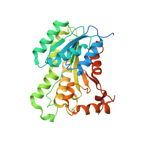
    Methicillin-resistant Staphylococcus aureus (MRSA) infections constitute a serious health threat worldwide, and novel antibiotics are therefore urgently needed. The enoyl-ACP reductase (saFabI) is essential for the S. aureus fatty acid biosynthesis and, hence, serves as an attractive drug target. We have obtained a series of snapshots of this enzyme that provide a mechanistic picture of ligand and inhibitor binding, including a dimer-tetramer transition combined with extensive conformational changes. Significantly, our results reveal key differences in ligand binding and recognition compared to orthologous proteins. The remarkable observed protein flexibility rationalizes our finding that saFabI is capable of efficiently reducing branched-chain fatty acid precursors. Importantly, branched-chain fatty acids represent a major fraction of the S. aureus cell membrane and are crucial for its in vivo fitness. Our discovery thus addresses a long-standing controversy regarding the essentiality of the fatty acid biosynthesis pathway in S. aureus rationalizing saFabI as a drug target.


  • Organizational Affiliation
    • Rudolf Virchow Center for Experimental Biomedicine, Institute for Structural Biology, University of Würzburg, D-97080 Würzburg, Germany.

Macromolecule Content 

  • Total Structure Weight: 126.37 kDa 
  • Atom Count: 8,101 
  • Modeled Residue Count: 1,018 
  • Deposited Residue Count: 1,108 
  • Unique protein chains: 1

Macromolecules

Find similar proteins by:|  3D Structure
Entity ID: 1
MoleculeChains  Sequence LengthOrganismDetailsImage
ENOYL-[ACYL-CARRIER-PROTEIN] REDUCTASE [NADPH]
A, B, C, D
277Staphylococcus aureusMutation(s): 0 
EC: 1.3.1.10

Small Molecules

Ligands 2 Unique
IDChains Name / Formula / InChI Key2D Diagram3D Interactions
NAP

Query on NAP


Download:Ideal Coordinates CCD File
F [auth A],
G [auth B],
I [auth C],
L [auth D]
NADP NICOTINAMIDE-ADENINE-DINUCLEOTIDE PHOSPHATE
C21 H28 N7 O17 P3
XJLXINKUBYWONI-NNYOXOHSSA-N
TCL

Query on TCL


Download:Ideal Coordinates CCD File
E [auth A],
H [auth B],
J [auth C],
K [auth D]
TRICLOSAN
C12 H7 Cl3 O2
XEFQLINVKFYRCS-UHFFFAOYSA-N
Binding Affinity Annotations 
IDSourceBinding Affinity
TCL BindingDB:  4ALL Ki: min: 0.05, max: 5 (nM) from 2 assay(s)
IC50: min: 59, max: 1100 (nM) from 4 assay(s)

Experimental Data & Validation

Experimental Data

  • Method: X-RAY DIFFRACTION
  • Resolution: 2.80 Å
  • R-Value Free:  0.262 (Depositor), 0.262 (DCC) 
  • R-Value Work:  0.214 (Depositor), 0.213 (DCC) 
  • R-Value Observed: 0.216 (Depositor) 
Space Group: P 21 21 21
Unit Cell:
Length ( Å )Angle ( ˚ )
a = 83.52α = 90
b = 111.91β = 90
c = 111.64γ = 90
Software Package:
Software NamePurpose
REFMACrefinement
iMOSFLMdata reduction
SCALAdata scaling
PHASERphasing

Structure Validation

View Full Validation Report


Ligand Structure Quality Assessment 


Entry History 

Deposition Data

Revision History  (Full details and data files)

  • Version 1.0: 2012-05-09
    Type: Initial release
  • Version 1.1: 2012-05-23
    Changes: Other
  • Version 1.2: 2023-12-20
    Changes: Data collection, Database references, Derived calculations, Other, Refinement description