4A6F | pdb_00004a6f

Crystal structure of Slm1-PH domain in complex with Phosphoserine


Experimental Data Snapshot

  • Method: X-RAY DIFFRACTION
  • Resolution: 1.68 Å
  • R-Value Free: 
    0.242 (Depositor), 0.252 (DCC) 
  • R-Value Work: 
    0.214 (Depositor), 0.239 (DCC) 
  • R-Value Observed: 
    0.215 (Depositor) 

Starting Model: experimental
View more details

wwPDB Validation 3D Report Full Report

Validation slider image for 4A6F

Ligand Structure Quality Assessment 


This is version 1.3 of the entry. See complete history

Literature

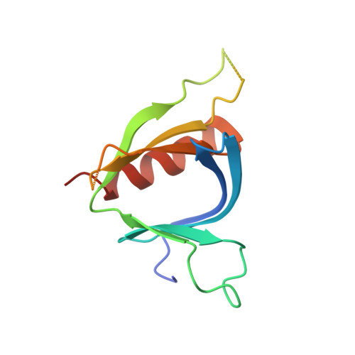
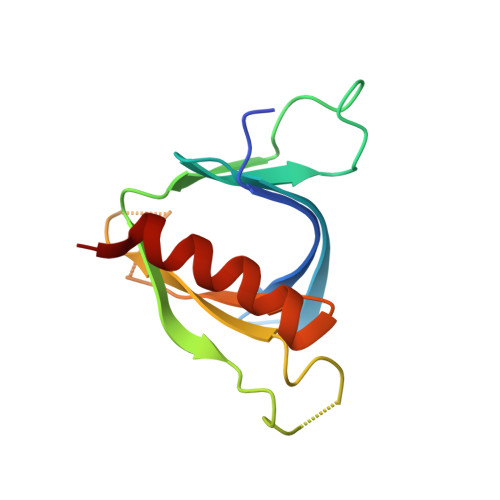
Structural Analyses of Slm1-Ph Domain Demonstrate Ligand Binding in the Non-Canonical Site

Anand, K.Maeda, K.Gavin, A.C.

(2012) PLoS One 7: 36526

  • DOI: https://doi.org/10.1371/journal.pone.0036526
  • Primary Citation Related Structures: 
    4A5K, 4A6F, 4A6H, 4A6K

  • PubMed Abstract: 

    Pleckstrin homology (PH) domains are common membrane-targeting modules and their best characterized ligands are a set of important signaling lipids that include phosphatidylinositol phosphates (PtdInsPs). PH domains recognize PtdInsPs through two distinct mechanisms that use different binding pockets on opposite sides of the β-strands 1 and 2: i) a canonical binding site delimited by the β1-β2 and β3-β4loops and ii) a non-canonical binding site bordered by the β1-β2 and β5-β6loops. The PH domain-containing protein Slm1 from budding yeast Saccharomyces cerevisiae is required for actin cytoskeleton polarization and cell growth. We recently reported that this PH domain binds PtdInsPs and phosphorylated sphingolipids in a cooperative manner. To study the structural basis for the Slm1-PH domain (Slm1-PH) specificity, we co-crystallized this domain with different soluble compounds that have structures analogous to anionic lipid head groups of reported Slm1 ligands: inositol 4-phosphate, which mimics phosphatidylinositol-4-phosphate (PtdIns(4)P), and phosphoserine as a surrogate for dihydrosphingosine 1-phosphate (DHS1-P). We found electron densities for the ligands within the so-called non-canonical binding site. An additional positively charged surface that contacts a phosphate group was identified next to the canonical binding site. Our results suggest that Slm1-PH utilizes a non-canonical binding site to bind PtdInsPs, similar to that described for the PH domains of β-spectrin, Tiam1 and ArhGAP9. Additionally, Slm1-PH may have retained an active canonical site. We propose that the presence of both a canonical and a non-canonical binding pocket in Slm1-PH may account for the cooperative binding to PtdInsPs and DHS-1P.


  • Organizational Affiliation
    • European Molecular Biology Laboratory Heidelberg, Structural and Computational Biology Unit, Heidelberg, Germany. anand@embl.de

Macromolecule Content 

  • Total Structure Weight: 28.46 kDa 
  • Atom Count: 1,962 
  • Modeled Residue Count: 215 
  • Deposited Residue Count: 240 
  • Unique protein chains: 2

Macromolecules

Find similar proteins by:|  3D Structure
Entity ID: 1
MoleculeChains  Sequence LengthOrganismDetailsImage
PHOSPHATIDYLINOSITOL 4,5-BISPHOSPHATE-BINDING PROTEIN SLM1120Saccharomyces cerevisiaeMutation(s): 0 
Membrane Entity: Yes 
UniProt
Find proteins for P40485 (Saccharomyces cerevisiae (strain ATCC 204508 / S288c))
Explore P40485 
Go to UniProtKB:  P40485
Entity Groups
Sequence Clusters30% Identity50% Identity70% Identity90% Identity95% Identity100% Identity
UniProt GroupP40485
Sequence Annotations
Expand
Reference Sequence
Find similar proteins by:|  3D Structure
Entity ID: 2
MoleculeChains  Sequence LengthOrganismDetailsImage
PHOSPHATIDYLINOSITOL 4,5-BISPHOSPHATE-BINDING PROTEIN SLM1120Saccharomyces cerevisiaeMutation(s): 0 
Membrane Entity: Yes 
UniProt
Find proteins for P40485 (Saccharomyces cerevisiae (strain ATCC 204508 / S288c))
Explore P40485 
Go to UniProtKB:  P40485
Entity Groups
Sequence Clusters30% Identity50% Identity70% Identity90% Identity95% Identity100% Identity
UniProt GroupP40485
Sequence Annotations
Expand
Reference Sequence

Experimental Data & Validation

Experimental Data

  • Method: X-RAY DIFFRACTION
  • Resolution: 1.68 Å
  • R-Value Free:  0.242 (Depositor), 0.252 (DCC) 
  • R-Value Work:  0.214 (Depositor), 0.239 (DCC) 
  • R-Value Observed: 0.215 (Depositor) 
Space Group: P 2 21 21
Unit Cell:
Length ( Å )Angle ( ˚ )
a = 37.54α = 90
b = 73.65β = 90
c = 82.47γ = 90
Software Package:
Software NamePurpose
PHENIXrefinement
XDSdata reduction
XSCALEdata scaling
PHASERphasing

Structure Validation

View Full Validation Report


Ligand Structure Quality Assessment 


Entry History 

Deposition Data

Revision History  (Full details and data files)

  • Version 1.0: 2012-06-13
    Type: Initial release
  • Version 1.1: 2013-02-06
    Changes: Atomic model
  • Version 1.2: 2019-10-16
    Changes: Data collection, Other
  • Version 1.3: 2023-12-20
    Changes: Data collection, Database references, Derived calculations, Refinement description