4A34 | pdb_00004a34

Crystal structure of the fucose mutarotase in complex with L-fucose from Streptococcus pneumoniae


Experimental Data Snapshot

  • Method: X-RAY DIFFRACTION
  • Resolution: 2.50 Å
  • R-Value Free: 
    0.292 (Depositor), 0.285 (DCC) 
  • R-Value Work: 
    0.225 (Depositor), 0.220 (DCC) 
  • R-Value Observed: 
    0.228 (Depositor) 

wwPDB Validation 3D Report Full Report

Validation slider image for 4A34

Ligand Structure Quality Assessment 


This is version 1.5 of the entry. See complete history

Literature

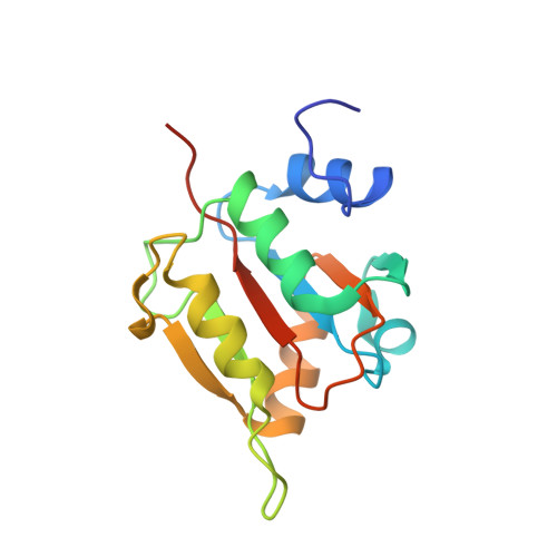
Structure of the Fucose Mutarotase from Streptococcus Pneumoniae in Complex with L-Fucose

Higgins, M.A.Boraston, A.B.

(2011) Acta Crystallogr Sect F Struct Biol Cryst Commun 67: 1524

  • DOI: https://doi.org/10.1107/S1744309111046343
  • Primary Citation Related Structures: 
    4A34

  • PubMed Abstract: 

    Streptococcus pneumoniae relies on a variety of carbohydrate-utilization pathways for both colonization of its human host and full virulence during the development of invasive disease. One such pathway is the fucose-utilization pathway, a component of which is fucose mutarotase (SpFcsU), an enzyme that performs the interconversion between α-L-fucose and β-L-fucose. This protein was crystallized and its three-dimensional structure was solved in complex with L-fucose. The structure shows a complex decameric quaternary structure with a high overall degree of structural identity to Escherichia coli FcsU (EcFcsU). Furthermore, the active-site architecture of SpFcsU is highly similar to that of EcFcsU. When considered in the context of the fucose-utilization pathway found in S. pneumoniae, SpFcsU appears to link the two halves of the pathway by enhancing the rate of conversion of the product of the final glycoside hydrolysis step, β-fucose, into the substrate for the fucose isomerase, α-fucose.


  • Organizational Affiliation
    • Department of Biochemistry and Microbiology, University of Victoria, Victoria, BC, Canada.

Macromolecule Content 

  • Total Structure Weight: 335.16 kDa 
  • Atom Count: 22,775 
  • Modeled Residue Count: 2,801 
  • Deposited Residue Count: 2,940 
  • Unique protein chains: 1

Macromolecules

Find similar proteins by:|  3D Structure
Entity ID: 1
MoleculeChains  Sequence LengthOrganismDetailsImage
RBSD/FUCU TRANSPORT PROTEIN FAMILY PROTEIN
A, B, C, D, E
A, B, C, D, E, F, G, H, I, J, K, L, M, N, O, P, Q, R, S, T
147Streptococcus pneumoniae TIGR4Mutation(s): 0 
UniProt
Find proteins for A0A0H2ZNZ5 (Streptococcus pneumoniae serotype 2 (strain D39 / NCTC 7466))
Explore A0A0H2ZNZ5 
Go to UniProtKB:  A0A0H2ZNZ5
Entity Groups
Sequence Clusters30% Identity50% Identity70% Identity90% Identity95% Identity100% Identity
UniProt GroupA0A0H2ZNZ5
Sequence Annotations
Expand
Reference Sequence

Small Molecules

Ligands 2 Unique
IDChains Name / Formula / InChI Key2D Diagram3D Interactions
FUL

Query on FUL


Download:Ideal Coordinates CCD File
BA [auth E]
DA [auth F]
EA [auth G]
FA [auth H]
GA [auth I]
BA [auth E],
DA [auth F],
EA [auth G],
FA [auth H],
GA [auth I],
HA [auth J],
JA [auth K],
LA [auth L],
NA [auth M],
OA [auth N],
QA [auth O],
RA [auth P],
SA [auth Q],
U [auth A],
UA [auth R],
V [auth B],
VA [auth S],
X [auth C],
XA [auth T],
Z [auth D]
beta-L-fucopyranose
C6 H12 O5
SHZGCJCMOBCMKK-KGJVWPDLSA-N
K

Query on K


Download:Ideal Coordinates CCD File
AA [auth D]
CA [auth E]
IA [auth J]
KA [auth K]
MA [auth L]
AA [auth D],
CA [auth E],
IA [auth J],
KA [auth K],
MA [auth L],
PA [auth N],
TA [auth Q],
W [auth B],
WA [auth S],
Y [auth C]
POTASSIUM ION
K
NPYPAHLBTDXSSS-UHFFFAOYSA-N

Experimental Data & Validation

Experimental Data

  • Method: X-RAY DIFFRACTION
  • Resolution: 2.50 Å
  • R-Value Free:  0.292 (Depositor), 0.285 (DCC) 
  • R-Value Work:  0.225 (Depositor), 0.220 (DCC) 
  • R-Value Observed: 0.228 (Depositor) 
Space Group: P 1 21 1
Unit Cell:
Length ( Å )Angle ( ˚ )
a = 64.319α = 90
b = 144.158β = 97.51
c = 165.46γ = 90
Software Package:
Software NamePurpose
REFMACrefinement

Structure Validation

View Full Validation Report


Ligand Structure Quality Assessment 


Entry History 

Deposition Data

Revision History  (Full details and data files)

  • Version 1.0: 2011-10-12
    Type: Initial release
  • Version 1.1: 2011-12-14
    Changes: Database references
  • Version 1.2: 2012-03-28
    Changes: Other
  • Version 1.3: 2013-09-04
    Changes: Atomic model, Derived calculations
  • Version 1.4: 2020-07-29
    Type: Remediation
    Reason: Carbohydrate remediation
    Changes: Data collection, Derived calculations, Other, Structure summary
  • Version 1.5: 2024-05-08
    Changes: Data collection, Database references, Structure summary