4ZZB | pdb_00004zzb

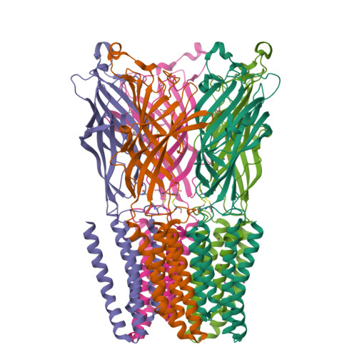
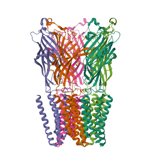
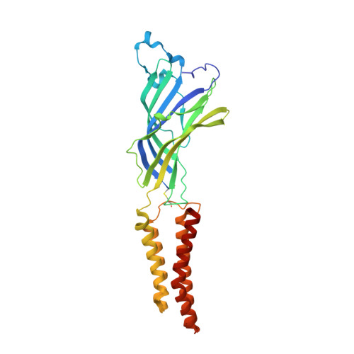
The GLIC pentameric Ligand-Gated Ion Channel Locally-closed form complexed to xenon


Experimental Data Snapshot

  • Method: X-RAY DIFFRACTION
  • Resolution: 3.40 Å
  • R-Value Free: 
    0.223 (Depositor), 0.240 (DCC) 
  • R-Value Work: 
    0.216 (Depositor), 0.250 (DCC) 
  • R-Value Observed: 
    0.217 (Depositor) 

wwPDB Validation   3D Report Full Report


Ligand Structure Quality Assessment 

Created with Raphaël 2.3.0Worse 01 BetterLigand structure goodness of fit to experimental dataBest fitted LMTClick on this verticalbar to view details

This is version 1.3 of the entry. See complete history


Literature

Structural Basis for Xenon Inhibition in a Cationic Pentameric Ligand-Gated Ion Channel.

Sauguet, L.Fourati, Z.Prange, T.Delarue, M.Colloc'h, N.

(2016) PLoS One 11: e0149795-e0149795

  • DOI: https://doi.org/10.1371/journal.pone.0149795
  • Primary Citation of Related Structures:  
    4ZZB, 4ZZC

  • PubMed Abstract: 

    GLIC receptor is a bacterial pentameric ligand-gated ion channel whose action is inhibited by xenon. Xenon has been used in clinical practice as a potent gaseous anaesthetic for decades, but the molecular mechanism of interactions with its integral membrane receptor targets remains poorly understood. Here we characterize by X-ray crystallography the xenon-binding sites within both the open and "locally-closed" (inactive) conformations of GLIC. Major binding sites of xenon, which differ between the two conformations, were identified in three distinct regions that all belong to the trans-membrane domain of GLIC: 1) in an intra-subunit cavity, 2) at the interface between adjacent subunits, and 3) in the pore. The pore site is unique to the locally-closed form where the binding of xenon effectively seals the channel. A putative mechanism of the inhibition of GLIC by xenon is proposed, which might be extended to other pentameric cationic ligand-gated ion channels.


  • Organizational Affiliation

    Unité de Dynamique Structurale des Macromolécules (UMR 3528 CNRS) Institut Pasteur, Paris, France.


Macromolecules
Find similar proteins by:  (by identity cutoff)  |  3D Structure
Entity ID: 1
MoleculeChains Sequence LengthOrganismDetailsImage
Proton-gated ion channel
A, B, C, D, E
317Gloeobacter violaceus PCC 8105Mutation(s): 3 
Gene Names: glvIglr4197
Membrane Entity: Yes 
UniProt
Find proteins for Q7NDN8 (Gloeobacter violaceus (strain ATCC 29082 / PCC 7421))
Explore Q7NDN8 
Go to UniProtKB:  Q7NDN8
Entity Groups  
Sequence Clusters30% Identity50% Identity70% Identity90% Identity95% Identity100% Identity
UniProt GroupQ7NDN8
Sequence Annotations
Expand
  • Reference Sequence
Small Molecules
Ligands 5 Unique
IDChains Name / Formula / InChI Key2D Diagram3D Interactions
LMT
Query on LMT

Download Ideal Coordinates CCD File 
F [auth A]DODECYL-BETA-D-MALTOSIDE
C24 H46 O11
NLEBIOOXCVAHBD-QKMCSOCLSA-N
XE
Query on XE

Download Ideal Coordinates CCD File 
AA [auth C]
BA [auth C]
CA [auth C]
DA [auth C]
HA [auth D]
XENON
Xe
FHNFHKCVQCLJFQ-UHFFFAOYSA-N
ACT
Query on ACT

Download Ideal Coordinates CCD File 
GA [auth D]
I [auth A]
NA [auth E]
R [auth B]
W [auth C]
ACETATE ION
C2 H3 O2
QTBSBXVTEAMEQO-UHFFFAOYSA-M
CL
Query on CL

Download Ideal Coordinates CCD File 
EA [auth D],
G [auth A],
LA [auth E],
P [auth B],
X [auth C]
CHLORIDE ION
Cl
VEXZGXHMUGYJMC-UHFFFAOYSA-M
NA
Query on NA

Download Ideal Coordinates CCD File 
FA [auth D],
H [auth A],
MA [auth E],
Q [auth B],
Y [auth C]
SODIUM ION
Na
FKNQFGJONOIPTF-UHFFFAOYSA-N
Experimental Data & Validation

Experimental Data

  • Method: X-RAY DIFFRACTION
  • Resolution: 3.40 Å
  • R-Value Free:  0.223 (Depositor), 0.240 (DCC) 
  • R-Value Work:  0.216 (Depositor), 0.250 (DCC) 
  • R-Value Observed: 0.217 (Depositor) 
Space Group: C 1 2 1
Unit Cell:
Length ( Å )Angle ( ˚ )
a = 176.966α = 90
b = 130.528β = 100.33
c = 157.526γ = 90
Software Package:
Software NamePurpose
BUSTERrefinement
XDSdata reduction
SCALAdata scaling
REFMACphasing

Structure Validation

View Full Validation Report


Ligand Structure Quality Assessment 

Created with Raphaël 2.3.0Worse 01 BetterLigand structure goodness of fit to experimental dataBest fitted LMTClick on this verticalbar to view details

Entry History 

Deposition Data

Revision History  (Full details and data files)

  • Version 1.0: 2016-03-02
    Type: Initial release
  • Version 1.1: 2016-03-09
    Changes: Database references
  • Version 1.2: 2018-11-21
    Changes: Data collection, Source and taxonomy
  • Version 1.3: 2024-11-13
    Changes: Data collection, Database references, Structure summary