4ZYB | pdb_00004zyb

High resolution structure of M23 peptidase LytM with substrate analogue


Experimental Data Snapshot

  • Method: X-RAY DIFFRACTION
  • Resolution: 1.50 Å
  • R-Value Free: 
    0.185 (Depositor), 0.183 (DCC) 
  • R-Value Work: 
    0.150 (Depositor), 0.149 (DCC) 
  • R-Value Observed: 
    0.151 (Depositor) 

Starting Model: experimental
View more details

wwPDB Validation 3D Report Full Report

Validation slider image for 4ZYB

Ligand Structure Quality Assessment 


This is version 1.2 of the entry. See complete history

Literature

High resolution structure of an M23 peptidase with a substrate analogue.

Grabowska, M.Jagielska, E.Czapinska, H.Bochtler, M.Sabala, I.

(2015) Sci Rep 5: 14833-14833

  • DOI: https://doi.org/10.1038/srep14833
  • Primary Citation Related Structures: 
    4ZYB

  • PubMed Abstract: 

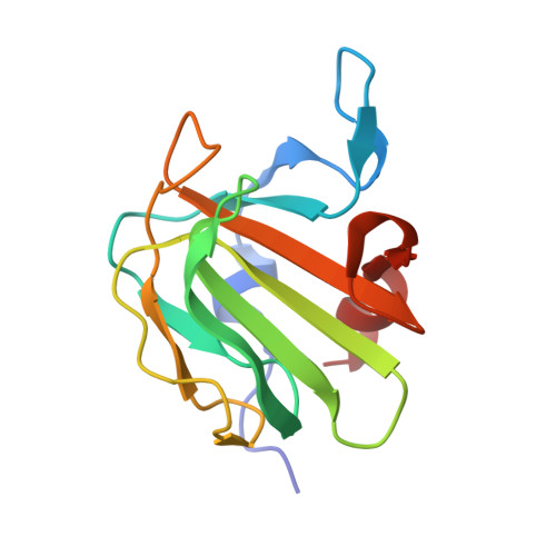
    LytM is a Staphylococcus aureus autolysin and a homologue of the S. simulans lysostaphin. Both enzymes are members of M23 metallopeptidase family (MEROPS) comprising primarily bacterial peptidoglycan hydrolases. LytM occurs naturally in a latent form, but can be activated by cleavage of an inhibitory N-terminal proregion. Here, we present a 1.45 Å crystal structure of LytM catalytic domain with a transition state analogue, tetraglycine phosphinate, bound in the active site. In the electron density, the active site of the peptidase, the phosphinate and the "diglycine" fragment on the P1' side of the transition state analogue are very well defined. The density is much poorer or even absent for the P1 side of the ligand. The structure is consistent with the involvement of His260 and/or His291 in the activation of the water nucleophile and suggests a possible catalytic role for Tyr204, which we confirmed by mutagenesis. Possible mechanisms of catalysis and the structural basis of substrate specificity are discussed based on the structure analysis.


  • Organizational Affiliation
    • International Institute of Molecular and Cell Biology, Ks. Trojdena 4, 02-109 Warsaw, Poland.

Macromolecule Content 

  • Total Structure Weight: 61.07 kDa 
  • Atom Count: 5,713 
  • Modeled Residue Count: 531 
  • Deposited Residue Count: 536 
  • Unique protein chains: 1

Macromolecules

Find similar proteins by:|  3D Structure
Entity ID: 1
MoleculeChains  Sequence LengthOrganismDetailsImage
Glycyl-glycine endopeptidase LytM
A, B, C, D
134Staphylococcus aureus subsp. aureus NCTC 8325Mutation(s): 0 
Gene Names: lytMSAS0252
EC: 3.4.24.75
UniProt
Find proteins for O33599 (Staphylococcus aureus (strain NCTC 8325 / PS 47))
Explore O33599 
Go to UniProtKB:  O33599
Entity Groups
Sequence Clusters30% Identity50% Identity70% Identity90% Identity95% Identity100% Identity
UniProt GroupO33599
Sequence Annotations
Expand
Reference Sequence

Small Molecules

Ligands 8 Unique
IDChains Name / Formula / InChI Key2D Diagram3D Interactions
4SQ

Query on 4SQ


Download:Ideal Coordinates CCD File
CA [auth D],
M [auth A],
T [auth B],
X [auth C]
tetraglycine phosphinate
C8 H16 N3 O6 P
UENGEQFXYOYUEI-UHFFFAOYSA-N
EPE

Query on EPE


Download:Ideal Coordinates CCD File
N [auth A],
Y [auth C]
4-(2-HYDROXYETHYL)-1-PIPERAZINE ETHANESULFONIC ACID
C8 H18 N2 O4 S
JKMHFZQWWAIEOD-UHFFFAOYSA-N
PEG

Query on PEG


Download:Ideal Coordinates CCD File
R [auth B]DI(HYDROXYETHYL)ETHER
C4 H10 O3
MTHSVFCYNBDYFN-UHFFFAOYSA-N
ZN

Query on ZN


Download:Ideal Coordinates CCD File
E [auth A],
O [auth B],
U [auth C],
Z [auth D]
ZINC ION
Zn
PTFCDOFLOPIGGS-UHFFFAOYSA-N
EDO

Query on EDO


Download:Ideal Coordinates CCD File
L [auth A],
S [auth B]
1,2-ETHANEDIOL
C2 H6 O2
LYCAIKOWRPUZTN-UHFFFAOYSA-N
CA

Query on CA


Download:Ideal Coordinates CCD File
AA [auth D],
F [auth A],
P [auth B],
V [auth C]
CALCIUM ION
Ca
BHPQYMZQTOCNFJ-UHFFFAOYSA-N
CL

Query on CL


Download:Ideal Coordinates CCD File
BA [auth D],
G [auth A],
H [auth A],
Q [auth B]
CHLORIDE ION
Cl
VEXZGXHMUGYJMC-UHFFFAOYSA-M
UNL

Query on UNL


Download:Ideal Coordinates CCD File
I [auth A],
J [auth A],
K [auth A],
W [auth C]
Unknown ligand
VEXZGXHMUGYJMC-UHFFFAOYSA-M

Experimental Data & Validation

Experimental Data

  • Method: X-RAY DIFFRACTION
  • Resolution: 1.50 Å
  • R-Value Free:  0.185 (Depositor), 0.183 (DCC) 
  • R-Value Work:  0.150 (Depositor), 0.149 (DCC) 
  • R-Value Observed: 0.151 (Depositor) 
Space Group: P 1 21 1
Unit Cell:
Length ( Å )Angle ( ˚ )
a = 31.988α = 90
b = 101.563β = 91.48
c = 77.763γ = 90
Software Package:
Software NamePurpose
PHENIXrefinement
XDSdata reduction
XDSdata scaling
MOLREPphasing
ARPmodel building

Structure Validation

View Full Validation Report


Ligand Structure Quality Assessment 


Entry History 

& Funding Information

Deposition Data


Funding OrganizationLocationGrant Number
NCBiRPolandPBS1/A8/8/2012

Revision History  (Full details and data files)

  • Version 1.0: 2015-10-21
    Type: Initial release
  • Version 1.1: 2018-04-25
    Changes: Data collection
  • Version 1.2: 2024-01-10
    Changes: Data collection, Database references, Derived calculations, Refinement description