4ZKD | pdb_00004zkd

Crystal structure of the S. cerevisiae Ski7 GTPase-like domain, bound to GDP and inorganic phosphate.


Experimental Data Snapshot

  • Method: X-RAY DIFFRACTION
  • Resolution: 2.18 Å
  • R-Value Free: 
    0.246 (Depositor), 0.253 (DCC) 
  • R-Value Work: 
    0.217 (Depositor), 0.226 (DCC) 
  • R-Value Observed: 
    0.219 (Depositor) 

wwPDB Validation 3D Report Full Report

Validation slider image for 4ZKD

Ligand Structure Quality Assessment 


This is version 1.3 of the entry. See complete history

Literature

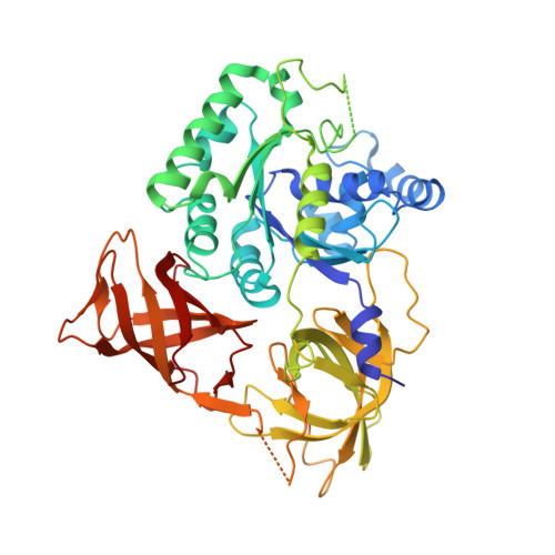
Saccharomyces cerevisiae Ski7 Is a GTP-Binding Protein Adopting the Characteristic Conformation of Active Translational GTPases.

Kowalinski, E.Schuller, A.Green, R.Conti, E.

(2015) Structure 23: 1336-1343

  • DOI: https://doi.org/10.1016/j.str.2015.04.018
  • Primary Citation Related Structures: 
    4ZKD, 4ZKE

  • PubMed Abstract: 

    Ski7 is a cofactor of the cytoplasmic exosome in budding yeast, functioning in both mRNA turnover and non-stop decay (NSD), a surveillance pathway that degrades faulty mRNAs lacking a stop codon. The C-terminal region of Ski7 (Ski7C) shares overall sequence similarity with the translational GTPase (trGTPase) Hbs1, but whether Ski7 has retained the properties of a trGTPase is unclear. Here, we report the high-resolution structures of Ski7C bound to either intact guanosine triphosphate (GTP) or guanosine diphosphate-Pi. The individual domains of Ski7C adopt the conformation characteristic of active trGTPases. Furthermore, the nucleotide-binding site of Ski7C shares similar features compared with active trGTPases, notably the presence of a characteristic monovalent cation. However, a suboptimal polar residue at the putative catalytic site and an unusual polar residue that interacts with the γ-phosphate of GTP distinguish Ski7 from other trGTPases, suggesting it might function rather as a GTP-binding protein than as a GTP-hydrolyzing enzyme.


  • Organizational Affiliation
    • Department of Structural Cell Biology Department, Max Planck Institute of Biochemistry, Am Klopferspitz 18, 82152 Martinsried, Germany.

Macromolecule Content 

  • Total Structure Weight: 57.22 kDa 
  • Atom Count: 3,853 
  • Modeled Residue Count: 470 
  • Deposited Residue Count: 499 
  • Unique protein chains: 1

Macromolecules

Find similar proteins by:|  3D Structure
Entity ID: 1
MoleculeChains  Sequence LengthOrganismDetailsImage
Superkiller protein 7499Saccharomyces cerevisiaeMutation(s): 0 
Gene Names: SKI7YOR076CYOR29-27
UniProt
Find proteins for Q08491 (Saccharomyces cerevisiae (strain ATCC 204508 / S288c))
Explore Q08491 
Go to UniProtKB:  Q08491
Entity Groups
Sequence Clusters30% Identity50% Identity70% Identity90% Identity95% Identity100% Identity
UniProt GroupQ08491
Sequence Annotations
Expand
Reference Sequence

Experimental Data & Validation

Experimental Data

  • Method: X-RAY DIFFRACTION
  • Resolution: 2.18 Å
  • R-Value Free:  0.246 (Depositor), 0.253 (DCC) 
  • R-Value Work:  0.217 (Depositor), 0.226 (DCC) 
  • R-Value Observed: 0.219 (Depositor) 
Space Group: C 2 2 21
Unit Cell:
Length ( Å )Angle ( ˚ )
a = 93.136α = 90
b = 120.617β = 90
c = 105.269γ = 90
Software Package:
Software NamePurpose
PHENIXrefinement
XDSdata reduction
XDSdata scaling
SHARPphasing

Structure Validation

View Full Validation Report


Ligand Structure Quality Assessment 


Entry History 

Deposition Data

Revision History  (Full details and data files)

  • Version 1.0: 2015-06-17
    Type: Initial release
  • Version 1.1: 2015-06-24
    Changes: Database references
  • Version 1.2: 2015-07-15
    Changes: Database references
  • Version 1.3: 2024-05-08
    Changes: Data collection, Database references, Derived calculations