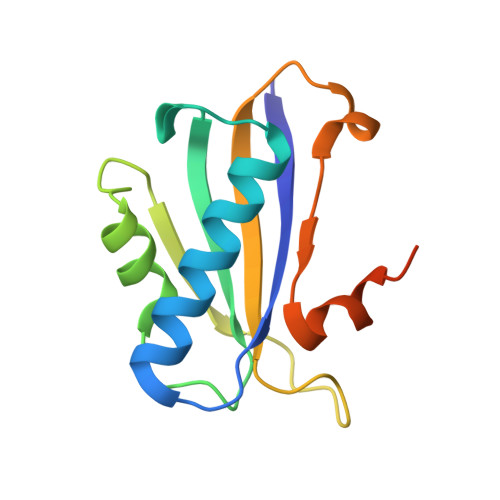
4ZK7 | pdb_00004zk7

Crystal structure of rescued two-component self-assembling tetrahedral cage T33-31


Experimental Data Snapshot

  • Method: X-RAY DIFFRACTION
  • Resolution: 3.40 Å
  • R-Value Free: 
    0.239 (Depositor), 0.228 (DCC) 
  • R-Value Work: 
    0.190 (Depositor) 
  • R-Value Observed: 
    0.192 (Depositor) 

wwPDB Validation 3D Report Full Report

Validation slider image for 4ZK7

This is version 1.3 of the entry. See complete history. 

Literature

Structure of a designed tetrahedral protein assembly variant engineered to have improved soluble expression.

Bale, J.B., Park, R.U., Liu, Y., Gonen, S., Gonen, T., Cascio, D., King, N.P., Yeates, T.O., Baker, D.

(2015) Protein Sci 24: 1695-1701

  • DOI: https://doi.org/10.1002/pro.2748
  • Primary Citation Related Structures: 
    4ZK7

  • PubMed Abstract: 

    We recently reported the development of a computational method for the design of coassembling multicomponent protein nanomaterials. While four such materials were validated at high-resolution by X-ray crystallography, low yield of soluble protein prevented X-ray structure determination of a fifth designed material, T33-09. Here we report the design and crystal structure of T33-31, a variant of T33-09 with improved soluble yield resulting from redesign efforts focused on mutating solvent-exposed side chains to charged amino acids. The structure is found to match the computational design model with atomic-level accuracy, providing further validation of the design approach and demonstrating a simple and potentially general means of improving the yield of designed protein nanomaterials.


  • Organizational Affiliation: 
    • Department of Biochemistry, University of Washington, Seattle, Washington, 98195.

Macromolecule Content 

  • Total Structure Weight: 324.48 kDa 
  • Atom Count: 20,678 
  • Modeled Residue Count: 2,646 
  • Deposited Residue Count: 2,892 
  • Unique protein chains: 2

Macromolecules

Find similar proteins by:|  3D Structure
Entity ID: 1
MoleculeChains  Sequence LengthOrganismDetailsImage
Chorismate mutase
A, B, C, D, E
A, B, C, D, E, F, G, H, I, J, K, L
130Thermus thermophilus HB8Mutation(s): 6 
Gene Names: TTHA0868
EC: 5.4.99.5
UniProt
Find proteins for Q5SJY4 (Thermus thermophilus (strain ATCC 27634 / DSM 579 / HB8))
Explore Q5SJY4 
Go to UniProtKB:  Q5SJY4
Entity Groups
Sequence Clusters30% Identity50% Identity70% Identity90% Identity95% Identity100% Identity
UniProt GroupQ5SJY4
Sequence Annotations
Expand
Reference Sequence
Find similar proteins by:|  3D Structure
Entity ID: 2
MoleculeChains  Sequence LengthOrganismDetailsImage
Divalent-cation tolerance protein CutA
M, N, O, P, Q
M, N, O, P, Q, R, S, T, U, V, W, X
111Thermus thermophilus HB8Mutation(s): 13 
Gene Names: cutA, TTHA1356
UniProt
Find proteins for Q7SIA8 (Thermus thermophilus (strain ATCC 27634 / DSM 579 / HB8))
Explore Q7SIA8 
Go to UniProtKB:  Q7SIA8
Entity Groups
Sequence Clusters30% Identity50% Identity70% Identity90% Identity95% Identity100% Identity
UniProt GroupQ7SIA8
Sequence Annotations
Expand
Reference Sequence

Experimental Data & Validation

Experimental Data

  • Method: X-RAY DIFFRACTION
  • Resolution: 3.40 Å
  • R-Value Free:  0.239 (Depositor), 0.228 (DCC) 
  • R-Value Work:  0.190 (Depositor) 
  • R-Value Observed: 0.192 (Depositor) 
Space Group: P 21 21 21
Unit Cell:
Length ( Å )Angle ( ˚ )
a = 121.1α = 90
b = 128.4β = 90
c = 204.7γ = 90
Software Package:
Software NamePurpose
XSCALEdata scaling
PHASERphasing
BUSTERrefinement
PDB_EXTRACTdata extraction
XDSdata reduction

Structure Validation

View Full Validation Report


Entry History 

Deposition Data

Revision History  (Full details and data files)

  • Version 1.0: 2015-07-29
    Type: Initial release
  • Version 1.1: 2015-10-07
    Changes: Database references
  • Version 1.2: 2017-11-01
    Changes: Author supporting evidence, Database references, Derived calculations
  • Version 1.3: 2024-03-06
    Changes: Data collection, Database references