4ZDM | pdb_00004zdm

Pleurobrachia bachei iGluR3 LBD Glycine Complex


Experimental Data Snapshot

  • Method: X-RAY DIFFRACTION
  • Resolution: 1.50 Å
  • R-Value Free: 
    0.177 (Depositor), 0.178 (DCC) 
  • R-Value Work: 
    0.156 (Depositor), 0.158 (DCC) 
  • R-Value Observed: 
    0.157 (Depositor) 

Starting Model: experimental
View more details

wwPDB Validation 3D Report Full Report

Validation slider image for 4ZDM

This is version 1.4 of the entry. See complete history

Literature

Glycine activated ion channel subunits encoded by ctenophore glutamate receptor genes.

Alberstein, R.Grey, R.Zimmet, A.Simmons, D.K.Mayer, M.L.

(2015) Proc Natl Acad Sci U S A 112: E6048-E6057

  • DOI: https://doi.org/10.1073/pnas.1513771112
  • Primary Citation Related Structures: 
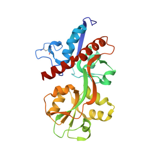
    4YKI, 4YKJ, 4YKK, 4YKP, 4ZDM

  • PubMed Abstract: 

    Recent genome projects for ctenophores have revealed the presence of numerous ionotropic glutamate receptors (iGluRs) in Mnemiopsis leidyi and Pleurobrachia bachei, among our earliest metazoan ancestors. Sequence alignments and phylogenetic analysis show that these form a distinct clade from the well-characterized AMPA, kainate, and NMDA iGluR subtypes found in vertebrates. Although annotated as glutamate and kainate receptors, crystal structures of the ML032222a and PbiGluR3 ligand-binding domains (LBDs) reveal endogenous glycine in the binding pocket, whereas ligand-binding assays show that glycine binds with nanomolar affinity; biochemical assays and structural analysis establish that glutamate is occluded from the binding cavity. Further analysis reveals ctenophore-specific features, such as an interdomain Arg-Glu salt bridge, present only in subunits that bind glycine, but also a conserved disulfide in loop 1 of the LBD that is found in all vertebrate NMDA but not AMPA or kainate receptors. We hypothesize that ctenophore iGluRs are related to an early ancestor of NMDA receptors, suggesting a common evolutionary path for ctenophores and bilaterian species, and suggest that future work should consider both glycine and glutamate as candidate neurotransmitters in ctenophore species.


  • Organizational Affiliation
    • Laboratory of Cellular and Molecular Neurophysiology, National Institute of Child Health and Human Development, National Institutes of Health, Bethesda, MD 20892;

Macromolecule Content 

  • Total Structure Weight: 29.15 kDa 
  • Atom Count: 2,448 
  • Modeled Residue Count: 249 
  • Deposited Residue Count: 261 
  • Unique protein chains: 1

Macromolecules

Find similar proteins by:|  3D Structure
Entity ID: 1
MoleculeChains  Sequence LengthOrganismDetailsImage
Glutamate receptor kainate-like protein261Pleurobrachia bacheiMutation(s): 0 
Gene Names: PbiGluR3
UniProt
Find proteins for H6S0J1 (Pleurobrachia bachei)
Explore H6S0J1 
Go to UniProtKB:  H6S0J1
Entity Groups
Sequence Clusters30% Identity50% Identity70% Identity90% Identity95% Identity100% Identity
UniProt GroupH6S0J1
Sequence Annotations
Expand
Reference Sequence

Experimental Data & Validation

Experimental Data

  • Method: X-RAY DIFFRACTION
  • Resolution: 1.50 Å
  • R-Value Free:  0.177 (Depositor), 0.178 (DCC) 
  • R-Value Work:  0.156 (Depositor), 0.158 (DCC) 
  • R-Value Observed: 0.157 (Depositor) 
Space Group: P 65 2 2
Unit Cell:
Length ( Å )Angle ( ˚ )
a = 159.85α = 90
b = 159.85β = 90
c = 55.614γ = 120
Software Package:
Software NamePurpose
PHENIXrefinement
HKL-2000data reduction
HKL-2000data scaling

Structure Validation

View Full Validation Report


Entry History 

Deposition Data

Revision History  (Full details and data files)

  • Version 1.0: 2015-10-21
    Type: Initial release
  • Version 1.1: 2015-10-28
    Changes: Database references
  • Version 1.2: 2015-11-18
    Changes: Database references
  • Version 1.3: 2023-09-27
    Changes: Data collection, Database references, Derived calculations, Refinement description
  • Version 1.4: 2024-10-16
    Changes: Structure summary