4ZAF | pdb_00004zaf

Structure of UbiX in complex with oxidised FMN and dimethylallyl monophosphate


Experimental Data Snapshot

  • Method: X-RAY DIFFRACTION
  • Resolution: 1.71 Å
  • R-Value Free: 
    0.172 (Depositor), 0.183 (DCC) 
  • R-Value Work: 
    0.137 (Depositor), 0.151 (DCC) 
  • R-Value Observed: 
    0.139 (Depositor) 

Starting Model: experimental
View more details

wwPDB Validation 3D Report Full Report

Validation slider image for 4ZAF

Ligand Structure Quality Assessment 


This is version 1.3 of the entry. See complete history

Literature

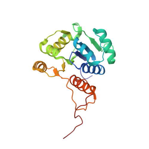
UbiX is a flavin prenyltransferase required for bacterial ubiquinone biosynthesis.

White, M.D.Payne, K.A.Fisher, K.Marshall, S.A.Parker, D.Rattray, N.J.Trivedi, D.K.Goodacre, R.Rigby, S.E.Scrutton, N.S.Hay, S.Leys, D.

(2015) Nature 522: 497-501

  • DOI: https://doi.org/10.1038/nature14559
  • Primary Citation Related Structures: 
    4ZAF, 4ZAG, 4ZAL, 4ZAN, 4ZAV, 4ZAW, 4ZAX, 4ZAY, 4ZAZ

  • PubMed Abstract: 

    Ubiquinone (also known as coenzyme Q) is a ubiquitous lipid-soluble redox cofactor that is an essential component of electron transfer chains. Eleven genes have been implicated in bacterial ubiquinone biosynthesis, including ubiX and ubiD, which are responsible for decarboxylation of the 3-octaprenyl-4-hydroxybenzoate precursor. Despite structural and biochemical characterization of UbiX as a flavin mononucleotide (FMN)-binding protein, no decarboxylase activity has been detected. Here we report that UbiX produces a novel flavin-derived cofactor required for the decarboxylase activity of UbiD. UbiX acts as a flavin prenyltransferase, linking a dimethylallyl moiety to the flavin N5 and C6 atoms. This adds a fourth non-aromatic ring to the flavin isoalloxazine group. In contrast to other prenyltransferases, UbiX is metal-independent and requires dimethylallyl-monophosphate as substrate. Kinetic crystallography reveals that the prenyltransferase mechanism of UbiX resembles that of the terpene synthases. The active site environment is dominated by π systems, which assist phosphate-C1' bond breakage following FMN reduction, leading to formation of the N5-C1' bond. UbiX then acts as a chaperone for adduct reorientation, via transient carbocation species, leading ultimately to formation of the dimethylallyl C3'-C6 bond. Our findings establish the mechanism for formation of a new flavin-derived cofactor, extending both flavin and terpenoid biochemical repertoires.


  • Organizational Affiliation
    • Centre for Synthetic Biology of Fine and Speciality Chemicals, Manchester Institute of Biotechnology, The University of Manchester, Manchester M1 7DN, UK.

Macromolecule Content 

  • Total Structure Weight: 23.28 kDa 
  • Atom Count: 1,814 
  • Modeled Residue Count: 207 
  • Deposited Residue Count: 209 
  • Unique protein chains: 1

Macromolecules

Find similar proteins by:|  3D Structure
Entity ID: 1
MoleculeChains  Sequence LengthOrganismDetailsImage
Probable aromatic acid decarboxylase209Pseudomonas aeruginosaMutation(s): 0 
Gene Names: PA4019
EC: 4.1.1 (PDB Primary Data), 2.5.1.129 (UniProt)
UniProt
Find proteins for Q9HX08 (Pseudomonas aeruginosa (strain ATCC 15692 / DSM 22644 / CIP 104116 / JCM 14847 / LMG 12228 / 1C / PRS 101 / PAO1))
Explore Q9HX08 
Go to UniProtKB:  Q9HX08
Entity Groups
Sequence Clusters30% Identity50% Identity70% Identity90% Identity95% Identity100% Identity
UniProt GroupQ9HX08
Sequence Annotations
Expand
Reference Sequence

Small Molecules

Ligands 4 Unique
IDChains Name / Formula / InChI Key2D Diagram3D Interactions
FMN

Query on FMN


Download:Ideal Coordinates CCD File
C [auth A]FLAVIN MONONUCLEOTIDE
C17 H21 N4 O9 P
FVTCRASFADXXNN-SCRDCRAPSA-N
4LR

Query on 4LR


Download:Ideal Coordinates CCD File
B [auth A]Dimethylallyl monophosphate
C5 H11 O4 P
MQCJHQBRIPSIKA-UHFFFAOYSA-N
SCN

Query on SCN


Download:Ideal Coordinates CCD File
D [auth A],
E [auth A],
F [auth A],
G [auth A]
THIOCYANATE ION
C N S
ZMZDMBWJUHKJPS-UHFFFAOYSA-M
K

Query on K


Download:Ideal Coordinates CCD File
H [auth A]POTASSIUM ION
K
NPYPAHLBTDXSSS-UHFFFAOYSA-N

Experimental Data & Validation

Experimental Data

  • Method: X-RAY DIFFRACTION
  • Resolution: 1.71 Å
  • R-Value Free:  0.172 (Depositor), 0.183 (DCC) 
  • R-Value Work:  0.137 (Depositor), 0.151 (DCC) 
  • R-Value Observed: 0.139 (Depositor) 
Space Group: F 2 3
Unit Cell:
Length ( Å )Angle ( ˚ )
a = 141.9α = 90
b = 141.9β = 90
c = 141.9γ = 90
Software Package:
Software NamePurpose
REFMACrefinement
xia2data reduction
xia2data scaling
REFMACphasing

Structure Validation

View Full Validation Report


Ligand Structure Quality Assessment 


Entry History 

& Funding Information

Deposition Data


Funding OrganizationLocationGrant Number
Biotechnology and Biological Sciences Research CouncilUnited Kingdom--

Revision History  (Full details and data files)

  • Version 1.0: 2015-06-17
    Type: Initial release
  • Version 1.1: 2015-07-01
    Changes: Database references
  • Version 1.2: 2017-08-30
    Changes: Author supporting evidence, Derived calculations
  • Version 1.3: 2024-05-01
    Changes: Data collection, Database references, Derived calculations, Refinement description