4Z9H | pdb_00004z9h

Asp-Tar from E. coli


Experimental Data Snapshot

  • Method: X-RAY DIFFRACTION
  • Resolution: 1.45 Å
  • R-Value Free: 
    0.205 (Depositor), 0.210 (DCC) 
  • R-Value Work: 
    0.173 (Depositor), 0.181 (DCC) 
  • R-Value Observed: 
    0.175 (Depositor) 

Starting Model: experimental
View more details

wwPDB Validation 3D Report Full Report

Validation slider image for 4Z9H

This is version 1.2 of the entry. See complete history

Literature

Structural Analysis of the Ligand-Binding Domain of the Aspartate Receptor Tar from Escherichia coli

Mise, T.

(2016) Biochemistry 55: 3708-3713

  • DOI: https://doi.org/10.1021/acs.biochem.6b00160
  • Primary Citation Related Structures: 
    4Z9H, 4Z9I, 4Z9J

  • PubMed Abstract: 

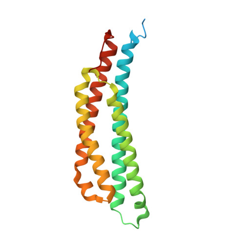
    The Escherichia coli cell-surface aspartate receptor Tar mediates bacterial chemotaxis toward an attractant, aspartate (Asp), and away from a repellent, Ni(2+). These signals are transmitted from the extracellular region of Tar to the cytoplasmic region via the transmembrane domain. The mechanism by which extracellular signals are transmitted into the cell through conformational changes in Tar is predicted to involve a piston displacement of one of the α4 helices of the homodimer. To understand the molecular mechanisms underlying the induction of Tar activity by an attractant, the three-dimensional structures of the E. coli Tar periplasmic domain with and without bound aspartate, Asp-Tar and apo-Tar, respectively, were determined. Of the two ligand-binding sites, only one site was occupied, and it clearly showed the electron density of an aspartate. The slight changes in conformation and the electrostatic surface potential around the aspartate-binding site were observed. In addition, the presence of an aspartate stabilized residues Phe-150' and Arg-73. A pistonlike displacement of helix α4b' was also induced by aspartate binding as predicted by the piston model. Taken together, these small changes might be related to the induction of Tar activity and might disturb binding of the second aspartate to the second binding site in E. coli.


  • Organizational Affiliation
    • 2-19-3 Misato, Okinawa-shi, Okinawa 904-2153, Japan.

Macromolecule Content 

  • Total Structure Weight: 44.7 kDa 
  • Atom Count: 2,835 
  • Modeled Residue Count: 323 
  • Deposited Residue Count: 392 
  • Unique protein chains: 1

Macromolecules

Find similar proteins by:|  3D Structure
Entity ID: 1
MoleculeChains  Sequence LengthOrganismDetailsImage
Methyl-accepting chemotaxis protein II
A, B
196Escherichia coli K-12Mutation(s): 0 
Gene Names: tarcheM
UniProt
Find proteins for P07017 (Escherichia coli (strain K12))
Explore P07017 
Go to UniProtKB:  P07017
Entity Groups
Sequence Clusters30% Identity50% Identity70% Identity90% Identity95% Identity100% Identity
UniProt GroupP07017
Sequence Annotations
Expand
Reference Sequence

Small Molecules

Ligands 1 Unique
IDChains Name / Formula / InChI Key2D Diagram3D Interactions
ASP

Query on ASP


Download:Ideal Coordinates CCD File
C [auth B]ASPARTIC ACID
C4 H7 N O4
CKLJMWTZIZZHCS-REOHCLBHSA-N

Experimental Data & Validation

Experimental Data

  • Method: X-RAY DIFFRACTION
  • Resolution: 1.45 Å
  • R-Value Free:  0.205 (Depositor), 0.210 (DCC) 
  • R-Value Work:  0.173 (Depositor), 0.181 (DCC) 
  • R-Value Observed: 0.175 (Depositor) 
Space Group: C 1 2 1
Unit Cell:
Length ( Å )Angle ( ˚ )
a = 81.32α = 90
b = 59.26β = 95.02
c = 79.23γ = 90
Software Package:
Software NamePurpose
REFMACrefinement
iMOSFLMdata reduction
SCALAdata scaling
MOLREPphasing

Structure Validation

View Full Validation Report


Entry History 

Deposition Data

  • Released Date: 2016-07-20 
  • Deposition Author(s): Mise, T.

Revision History  (Full details and data files)

  • Version 1.0: 2016-07-20
    Type: Initial release
  • Version 1.1: 2020-02-19
    Changes: Data collection, Database references, Derived calculations
  • Version 1.2: 2023-11-08
    Changes: Data collection, Database references, Refinement description