4Z9G | pdb_00004z9g

Crystal structure of human corticotropin-releasing factor receptor 1 (CRF1R) in complex with the antagonist CP-376395 in a hexagonal setting with translational non-crystallographic symmetry


Experimental Data Snapshot

  • Method: X-RAY DIFFRACTION
  • Resolution: 3.18 Å
  • R-Value Free: 
    0.289 (Depositor), 0.304 (DCC) 
  • R-Value Work: 
    0.244 (Depositor), 0.260 (DCC) 
  • R-Value Observed: 
    0.247 (Depositor) 

Starting Model: experimental
View more details

wwPDB Validation 3D Report Full Report

Validation slider image for 4Z9G

Ligand Structure Quality Assessment 


This is version 1.3 of the entry. See complete history

Literature

Decoding Corticotropin-Releasing Factor Receptor Type 1 Crystal Structures.

Dore, A.S.Bortolato, A.Hollenstein, K.Cheng, R.K.Y.Read, R.J.Marshall, F.H.

(2017) Curr Mol Pharmacol 10: 334-344

  • DOI: https://doi.org/10.2174/1874467210666170110114727
  • Primary Citation Related Structures: 
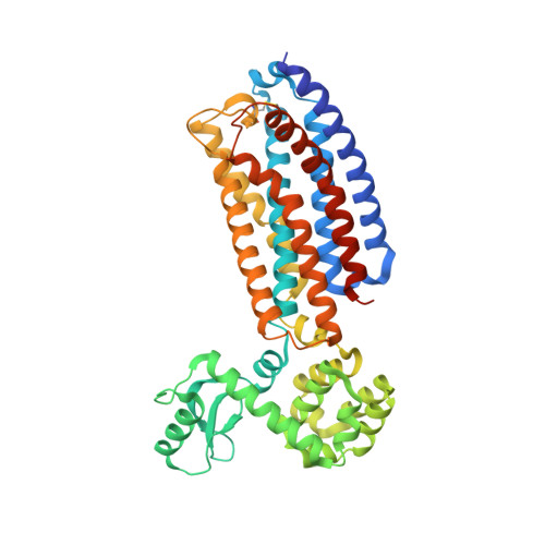
    4Z9G

  • PubMed Abstract: 

    The structural analysis of class B G protein-coupled receptors (GPCR), cell surface proteins responding to peptide hormones, has until recently been restricted to the extracellular domain (ECD). Corticotropin-releasing factor receptor type 1 (CRF1R) is a class B receptor mediating stress response and also considered a drug target for depression and anxiety. Here we report the crystal structure of the transmembrane domain of human CRF1R in complex with the small-molecule antagonist CP-376395 in a hexagonal setting with translational non-crystallographic symmetry. Molecular dynamics and metadynamics simulations on this novel structure and the existing TMD structure for CRF1R provides insight as to how the small molecule ligand gains access to the induced-fit allosteric binding site with implications for the observed selectivity against CRF2R. Furthermore, molecular dynamics simulations performed using a full-length receptor model point to key interactions between the ECD and extracellular loop 3 of the TMD providing insight into the full inactive state of multidomain class B GPCRs.


  • Organizational Affiliation
    • Heptares Therapeutics Ltd, BioPark, Broadwater Road, Welwyn Garden City, Herts, AL7 3AX. United Kingdom.

Macromolecule Content 

  • Total Structure Weight: 156.02 kDa 
  • Atom Count: 10,244 
  • Modeled Residue Count: 1,239 
  • Deposited Residue Count: 1,329 
  • Unique protein chains: 1

Macromolecules

Find similar proteins by:|  3D Structure
Entity ID: 1
MoleculeChains  Sequence LengthOrganismDetailsImage
Corticotropin-releasing factor receptor 1,Lysozyme,Corticotropin-releasing factor receptor 1
A, B, C
443Homo sapiensEnterobacteria phage RB51Mutation(s): 13 
Gene Names: CRHR1CRFRCRFR1CRHReRB51ORF131
EC: 3.2.1.17
Membrane Entity: Yes 
UniProt & NIH Common Fund Data Resources
Find proteins for C3V2B5 (Enterobacteria phage RB51)
Explore C3V2B5 
Go to UniProtKB:  C3V2B5
Find proteins for P34998 (Homo sapiens)
Explore P34998 
Go to UniProtKB:  P34998
PHAROS:  P34998
GTEx:  ENSG00000120088 
Entity Groups
Sequence Clusters30% Identity50% Identity70% Identity90% Identity95% Identity100% Identity
UniProt GroupsC3V2B5P34998
Sequence Annotations
Expand
Reference Sequence

Small Molecules

Ligands 3 Unique
IDChains Name / Formula / InChI Key2D Diagram3D Interactions
1Q5

Query on 1Q5


Download:Ideal Coordinates CCD File
G [auth A],
M [auth B],
R [auth C]
3,6-dimethyl-N-(pentan-3-yl)-2-(2,4,6-trimethylphenoxy)pyridin-4-amine
C21 H30 N2 O
VIZBSVDBNLAVAW-UHFFFAOYSA-N
OLA

Query on OLA


Download:Ideal Coordinates CCD File
D [auth A]
E [auth A]
F [auth A]
J [auth B]
K [auth B]
D [auth A],
E [auth A],
F [auth A],
J [auth B],
K [auth B],
L [auth B],
N [auth C],
O [auth C],
P [auth C],
Q [auth C]
OLEIC ACID
C18 H34 O2
ZQPPMHVWECSIRJ-KTKRTIGZSA-N
SO4

Query on SO4


Download:Ideal Coordinates CCD File
H [auth A],
I [auth A],
S [auth C]
SULFATE ION
O4 S
QAOWNCQODCNURD-UHFFFAOYSA-L
Binding Affinity Annotations 
IDSourceBinding Affinity
1Q5 BindingDB:  4Z9G IC50: 5.1 (nM) from 1 assay(s)

Experimental Data & Validation

Experimental Data

  • Method: X-RAY DIFFRACTION
  • Resolution: 3.18 Å
  • R-Value Free:  0.289 (Depositor), 0.304 (DCC) 
  • R-Value Work:  0.244 (Depositor), 0.260 (DCC) 
  • R-Value Observed: 0.247 (Depositor) 
Space Group: P 6
Unit Cell:
Length ( Å )Angle ( ˚ )
a = 189.359α = 90
b = 189.359β = 90
c = 88.575γ = 120
Software Package:
Software NamePurpose
Aimlessdata scaling
PHASERphasing
PHENIXrefinement
PDB_EXTRACTdata extraction

Structure Validation

View Full Validation Report


Ligand Structure Quality Assessment 


Entry History 

Deposition Data

Revision History  (Full details and data files)

  • Version 1.0: 2016-06-29
    Type: Initial release
  • Version 1.1: 2017-12-06
    Changes: Database references
  • Version 1.2: 2024-01-10
    Changes: Data collection, Database references, Refinement description
  • Version 1.3: 2024-11-13
    Changes: Structure summary