4Z79 | pdb_00004z79

Leiomodin-1 Actin-Binding Site 2 (ABS2)


Experimental Data Snapshot

  • Method: X-RAY DIFFRACTION
  • Resolution: 1.54 Å
  • R-Value Free: 
    0.177 (Depositor), 0.177 (DCC) 
  • R-Value Work: 
    0.156 (Depositor), 0.162 (DCC) 
  • R-Value Observed: 
    0.157 (Depositor) 

Starting Model: experimental
View more details

wwPDB Validation 3D Report Full Report

Validation slider image for 4Z79

This is version 1.4 of the entry. See complete history

Literature

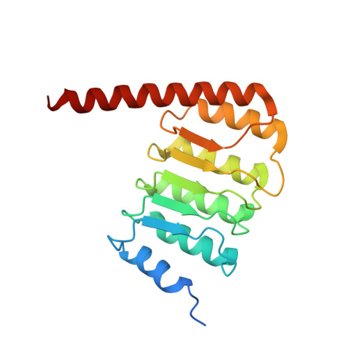
How Leiomodin and Tropomodulin use a common fold for different actin assembly functions.

Boczkowska, M.Rebowski, G.Kremneva, E.Lappalainen, P.Dominguez, R.

(2015) Nat Commun 6: 8314-8314

  • DOI: https://doi.org/10.1038/ncomms9314
  • Primary Citation Related Structures: 
    4Z79, 4Z8G, 4Z94

  • PubMed Abstract: 

    How proteins sharing a common fold have evolved different functions is a fundamental question in biology. Tropomodulins (Tmods) are prototypical actin filament pointed-end-capping proteins, whereas their homologues, Leiomodins (Lmods), are powerful filament nucleators. We show that Tmods and Lmods do not compete biochemically, and display similar but distinct localization in sarcomeres. Changes along the polypeptide chains of Tmods and Lmods exquisitely adapt their functions for capping versus nucleation. Tmods have alternating tropomyosin (TM)- and actin-binding sites (TMBS1, ABS1, TMBS2 and ABS2). Lmods additionally contain a C-terminal extension featuring an actin-binding WH2 domain. Unexpectedly, the different activities of Tmods and Lmods do not arise from the Lmod-specific extension. Instead, nucleation by Lmods depends on two major adaptations-the loss of pointed-end-capping elements present in Tmods and the specialization of the highly conserved ABS2 for recruitment of two or more actin subunits. The WH2 domain plays only an auxiliary role in nucleation.


  • Organizational Affiliation
    • Department of Physiology, Perelman School of Medicine, University of Pennsylvania, Philadelphia, Pennsylvania 19104, USA.

Macromolecule Content 

  • Total Structure Weight: 21.93 kDa 
  • Atom Count: 1,692 
  • Modeled Residue Count: 173 
  • Deposited Residue Count: 188 
  • Unique protein chains: 1

Macromolecules

Find similar proteins by:|  3D Structure
Entity ID: 1
MoleculeChains  Sequence LengthOrganismDetailsImage
Leiomodin-1 Actin-Binding Site 2 (ABS2)188Homo sapiensMutation(s): 0 
Gene Names: LMOD1
UniProt & NIH Common Fund Data Resources
Find proteins for P29536 (Homo sapiens)
Explore P29536 
Go to UniProtKB:  P29536
PHAROS:  P29536
GTEx:  ENSG00000163431 
Entity Groups
Sequence Clusters30% Identity50% Identity70% Identity90% Identity95% Identity100% Identity
UniProt GroupP29536
Sequence Annotations
Expand
Reference Sequence

Small Molecules

Ligands 3 Unique
IDChains Name / Formula / InChI Key2D Diagram3D Interactions
GOL

Query on GOL


Download:Ideal Coordinates CCD File
B [auth A]
C [auth A]
D [auth A]
E [auth A]
F [auth A]
B [auth A],
C [auth A],
D [auth A],
E [auth A],
F [auth A],
G [auth A]
GLYCEROL
C3 H8 O3
PEDCQBHIVMGVHV-UHFFFAOYSA-N
CL

Query on CL


Download:Ideal Coordinates CCD File
H [auth A]CHLORIDE ION
Cl
VEXZGXHMUGYJMC-UHFFFAOYSA-M
NA

Query on NA


Download:Ideal Coordinates CCD File
I [auth A],
J [auth A],
K [auth A]
SODIUM ION
Na
FKNQFGJONOIPTF-UHFFFAOYSA-N

Experimental Data & Validation

Experimental Data

  • Method: X-RAY DIFFRACTION
  • Resolution: 1.54 Å
  • R-Value Free:  0.177 (Depositor), 0.177 (DCC) 
  • R-Value Work:  0.156 (Depositor), 0.162 (DCC) 
  • R-Value Observed: 0.157 (Depositor) 
Space Group: P 21 21 21
Unit Cell:
Length ( Å )Angle ( ˚ )
a = 49.019α = 90
b = 52.192β = 90
c = 75.89γ = 90
Software Package:
Software NamePurpose
PHENIXrefinement
SAINTdata reduction
Cootmodel building
PHENIXphasing
SAINTdata scaling

Structure Validation

View Full Validation Report


Entry History 

& Funding Information

Deposition Data


Funding OrganizationLocationGrant Number
National Institutes of Health/National Institute of General Medical Sciences (NIH/NIGMS)United StatesR01 GM073791

Revision History  (Full details and data files)

  • Version 1.0: 2015-10-21
    Type: Initial release
  • Version 1.1: 2017-09-20
    Changes: Author supporting evidence, Derived calculations, Refinement description
  • Version 1.2: 2018-03-07
    Changes: Data collection
  • Version 1.3: 2019-12-25
    Changes: Author supporting evidence
  • Version 1.4: 2023-09-27
    Changes: Data collection, Database references, Derived calculations, Refinement description