4Z3C | pdb_00004z3c

Zinc finger region of human TET3 in complex with CpG DNA


Experimental Data Snapshot

  • Method: X-RAY DIFFRACTION
  • Resolution: 1.57 Å
  • R-Value Free: 
    0.253 (Depositor), 0.262 (DCC) 
  • R-Value Work: 
    0.215 (Depositor), 0.219 (DCC) 
  • R-Value Observed: 
    0.216 (Depositor) 

Starting Model: experimental
View more details

wwPDB Validation 3D Report Full Report

Validation slider image for 4Z3C

This is version 1.3 of the entry. See complete history

Literature

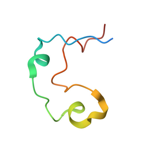
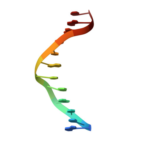
DNA Sequence Recognition of Human CXXC Domains and Their Structural Determinants.

Xu, C.Liu, K.Lei, M.Yang, A.Li, Y.Hughes, T.R.Min, J.

(2017) Structure 

  • DOI: https://doi.org/10.1016/j.str.2017.11.022
  • Primary Citation Related Structures: 
    4NW3, 4O64, 4PZI, 4Z3C, 5VC9, 5W9Q, 5W9S, 6ASB, 6ASD

  • PubMed Abstract: 

    The CXXC domain, first identified as the reader of unmodified CpG dinucleotide, plays important roles in epigenetic regulation by targeting various activities to CpG islands. Here we systematically measured and compared the DNA-binding selectivities of all known human CXXC domains by different binding assays, and complemented the existing function-based classification of human CXXC domains with a classification based on their DNA selectivities. Through a series of crystal structures of CXXC domains with DNA ligands, we unravel the molecular mechanisms of how these CXXC domains, including single CXXC domains and tandem CXXC-PHD domains, recognize distinct DNA ligands, which further supports our classification of human CXXC domains and also provides insights into selective recruitment of chromatin modifiers to their respective targets via CXXC domains recognizing different genomic DNA sequences. Our study facilitates the understanding of the relationship between the DNA-binding specificities of the CXXC proteins and their biological functions.


  • Organizational Affiliation
    • Structural Genomics Consortium, University of Toronto, Toronto, ON M5G 1L7, Canada.

Macromolecule Content 

  • Total Structure Weight: 13.35 kDa 
  • Atom Count: 936 
  • Modeled Residue Count: 69 
  • Deposited Residue Count: 76 
  • Unique protein chains: 1
  • Unique nucleic acid chains: 1

Macromolecules


Find similar proteins by:|  3D Structure
Entity ID: 2
MoleculeChains  Sequence LengthOrganismDetailsImage
Methylcytosine dioxygenase52Homo sapiensMutation(s): 0 
Gene Names: TET3
EC: 1.14.11.80
UniProt & NIH Common Fund Data Resources
Find proteins for O43151 (Homo sapiens)
Explore O43151 
Go to UniProtKB:  O43151
PHAROS:  O43151
GTEx:  ENSG00000187605 
Entity Groups
Sequence Clusters30% Identity50% Identity70% Identity90% Identity95% Identity100% Identity
UniProt GroupO43151
Sequence Annotations
Expand
Reference Sequence
Find similar nucleic acids by:  Sequence
Entity ID: 1
MoleculeChains LengthOrganismImage
DNA (5'-D(*GP*CP*CP*AP*AP*CP*GP*TP*TP*GP*GP*C)-3')
A, B
12synthetic construct
Sequence Annotations
Expand
Reference Sequence

Experimental Data & Validation

Experimental Data

  • Method: X-RAY DIFFRACTION
  • Resolution: 1.57 Å
  • R-Value Free:  0.253 (Depositor), 0.262 (DCC) 
  • R-Value Work:  0.215 (Depositor), 0.219 (DCC) 
  • R-Value Observed: 0.216 (Depositor) 
Space Group: P 1 21 1
Unit Cell:
Length ( Å )Angle ( ˚ )
a = 27.616α = 90
b = 54.744β = 103.53
c = 40.228γ = 90
Software Package:
Software NamePurpose
Aimlessdata scaling
REFMACrefinement
PDB_EXTRACTdata extraction
XDSdata scaling
PHASERphasing

Structure Validation

View Full Validation Report


Entry History 

Deposition Data

Revision History  (Full details and data files)

  • Version 1.0: 2015-04-29
    Type: Initial release
  • Version 1.1: 2017-11-22
    Changes: Advisory, Derived calculations, Refinement description, Source and taxonomy
  • Version 1.2: 2018-01-03
    Changes: Database references, Structure summary
  • Version 1.3: 2023-09-27
    Changes: Data collection, Database references, Refinement description