4Z2S | pdb_00004z2s

The crystal structure of Sclerotium Rolfsii lectin variant 2 (SSR2) in complex with N-acetyl-glucosamine


Experimental Data Snapshot

  • Method: X-RAY DIFFRACTION
  • Resolution: 1.70 Å
  • R-Value Free: 
    0.203 (Depositor), 0.209 (DCC) 
  • R-Value Work: 
    0.165 (Depositor), 0.176 (DCC) 
  • R-Value Observed: 
    0.167 (Depositor) 

Starting Model: experimental
View more details

wwPDB Validation 3D Report Full Report

Validation slider image for 4Z2S

Ligand Structure Quality Assessment 


This is version 2.1 of the entry. See complete history

Literature

Molecular Cloning, Carbohydrate Specificity and the Crystal Structure of Two Sclerotium rolfsii Lectin Variants.

Peppa, V.I.Venkat, H.Kantsadi, A.L.Inamdar, S.R.Bhat, G.G.Eligar, S.Shivanand, A.Chachadi, V.B.Satisha, G.J.Swamy, B.M.Skamnaki, V.T.Zographos, S.E.Leonidas, D.D.

(2015) Molecules 20: 10848-10865

  • DOI: https://doi.org/10.3390/molecules200610848
  • Primary Citation Related Structures: 
    4YLD, 4Z2F, 4Z2Q, 4Z2S

  • PubMed Abstract: 

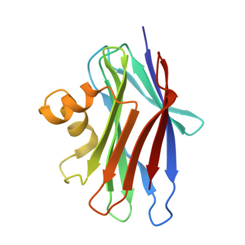
    SRL is a cell wall associated developmental-stage specific lectin secreted by Sclerotium rolfsii, a soil-born pathogenic fungus. SRL displays specificity for TF antigen (Galβ1→3GalNAc-α-Ser//Thr) expressed in all cancer types and has tumour suppressing effects in vivo. Considering the immense potential of SRL in cancer research, we have generated two variant gene constructs of SRL and expressed in E. coli to refine the sugar specificity and solubility by altering the surface charge. SSR1 and SSR2 are two different recombinant variants of SRL, both of which recognize TF antigen but only SSR1 binds to Tn antigen (GalNAcα-Ser/Thr). The glycan array analysis of the variants demonstrated that SSR1 recognizes TF antigen and their derivative with high affinity similar to SRL but showed highest affinity towards the sialylated Tn antigen, unlike SRL. The carbohydrate binding property of SSR2 remains unaltered compared to SRL. The crystal structures of the two variants were determined in free form and in complex with N-acetylglucosamine at 1.7 Å and 1.6 Å resolution, respectively. Structural analysis highlighted the structural basis of the fine carbohydrate specificity of the two SRL variants and results are in agreement with glycan array analysis.


  • Organizational Affiliation
    • Department of Biochemistry and Biotechnology, University of Thessaly, 26 Ploutonos Street, Larissa 41221, Greece. vikpeppa@gmail.com.

Macromolecule Content 

  • Total Structure Weight: 33.48 kDa 
  • Atom Count: 2,764 
  • Modeled Residue Count: 284 
  • Deposited Residue Count: 284 
  • Unique protein chains: 1

Macromolecules

Find similar proteins by:|  3D Structure
Entity ID: 1
MoleculeChains  Sequence LengthOrganismDetailsImage
Sclerotium Rolfsii lectin variant
A, B
142Agroathelia rolfsiiMutation(s): 0 
UniProt
Find proteins for A0A0M3KL31 (Agroathelia rolfsii)
Explore A0A0M3KL31 
Go to UniProtKB:  A0A0M3KL31
Entity Groups
Sequence Clusters30% Identity50% Identity70% Identity90% Identity95% Identity100% Identity
UniProt GroupA0A0M3KL31
Sequence Annotations
Expand
Reference Sequence

Small Molecules

Ligands 4 Unique
IDChains Name / Formula / InChI Key2D Diagram3D Interactions
NAG

Query on NAG


Download:Ideal Coordinates CCD File
E [auth A],
H [auth B]
2-acetamido-2-deoxy-beta-D-glucopyranose
C8 H15 N O6
OVRNDRQMDRJTHS-FMDGEEDCSA-N
NDG

Query on NDG


Download:Ideal Coordinates CCD File
D [auth A],
G [auth B]
2-acetamido-2-deoxy-alpha-D-glucopyranose
C8 H15 N O6
OVRNDRQMDRJTHS-PVFLNQBWSA-N
TRS

Query on TRS


Download:Ideal Coordinates CCD File
C [auth A]2-AMINO-2-HYDROXYMETHYL-PROPANE-1,3-DIOL
C4 H12 N O3
LENZDBCJOHFCAS-UHFFFAOYSA-O
MPD

Query on MPD


Download:Ideal Coordinates CCD File
F [auth B](4S)-2-METHYL-2,4-PENTANEDIOL
C6 H14 O2
SVTBMSDMJJWYQN-YFKPBYRVSA-N

Experimental Data & Validation

Experimental Data

  • Method: X-RAY DIFFRACTION
  • Resolution: 1.70 Å
  • R-Value Free:  0.203 (Depositor), 0.209 (DCC) 
  • R-Value Work:  0.165 (Depositor), 0.176 (DCC) 
  • R-Value Observed: 0.167 (Depositor) 
Space Group: P 42 21 2
Unit Cell:
Length ( Å )Angle ( ˚ )
a = 100.025α = 90
b = 100.025β = 90
c = 63.783γ = 90
Software Package:
Software NamePurpose
REFMACrefinement
CrysalisProdata reduction
SCALAdata scaling
PHENIXphasing

Structure Validation

View Full Validation Report


Ligand Structure Quality Assessment 


Entry History 

Deposition Data

Revision History  (Full details and data files)

  • Version 1.0: 2015-07-01
    Type: Initial release
  • Version 2.0: 2020-07-29
    Type: Remediation
    Reason: Carbohydrate remediation
    Changes: Atomic model, Data collection, Derived calculations, Structure summary
  • Version 2.1: 2024-01-10
    Changes: Data collection, Database references, Refinement description, Structure summary