4Z03 | pdb_00004z03

C. bescii Family 3 pectate lyase double mutant K108A in complex with trigalacturonic acid


Experimental Data Snapshot

  • Method: X-RAY DIFFRACTION
  • Resolution: 1.40 Å
  • R-Value Free: 
    0.184 (Depositor), 0.184 (DCC) 
  • R-Value Work: 
    0.122 (Depositor), 0.122 (DCC) 
  • R-Value Observed: 
    0.125 (Depositor) 

wwPDB Validation 3D Report Full Report

Validation slider image for 4Z03

Ligand Structure Quality Assessment 


This is version 2.1 of the entry. See complete history

Literature

The catalytic mechanism and unique low pH optimum of Caldicellulosiruptor bescii family 3 pectate lyase.

Alahuhta, M.Taylor, L.E.Brunecky, R.Sammond, D.W.Michener, W.Adams, M.W.Himmel, M.E.Bomble, Y.J.Lunin, V.

(2015) Acta Crystallogr D Biol Crystallogr 71: 1946-1954

  • DOI: https://doi.org/10.1107/S1399004715013760
  • Primary Citation Related Structures: 
    4YZ0, 4YZA, 4YZQ, 4YZX, 4Z03, 4Z05, 4Z06

  • PubMed Abstract: 

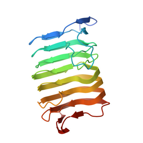
    The unique active site of the Caldicellulosiruptor bescii family 3 pectate lyase (PL3) enzyme has been thoroughly characterized using a series of point mutations, X-ray crystallography, pK(a) calculations and biochemical assays. The X-ray structures of seven PL3 active-site mutants, five of them in complex with intact trigalacturonic acid, were solved and characterized structurally, biochemically and computationally. The results confirmed that Lys108 is the catalytic base, but there is no clear candidate for the catalytic acid. However, the reaction mechanism can also be explained by an antiperiplanar trans-elimination reaction, in which Lys108 abstracts a proton from the C5 atom without the help of simultaneous proton donation by an acidic residue. An acidified water molecule completes the anti β-elimination reaction by protonating the O4 atom of the substrate. Both the C5 hydrogen and C4 hydroxyl groups of the substrate must be orientated in axial configurations, as for galacturonic acid, for this to be possible. The wild-type C. bescii PL3 displays a pH optimum that is lower than that of Bacillus subtilis PL1 according to activity measurements, indicating that C. bescii PL3 has acquired a lower pH optimum by utilizing lysine instead of arginine as the catalytic base, as well as by lowering the pK(a) of the catalytic base in a unique active-site environment.


  • Organizational Affiliation
    • BioSciences Center, National Renewable Energy Laboratory, 15013 Denver West Parkway, Golden, CO 80401, USA.

Macromolecule Content 

  • Total Structure Weight: 46.68 kDa 
  • Atom Count: 4,066 
  • Modeled Residue Count: 391 
  • Deposited Residue Count: 408 
  • Unique protein chains: 1

Macromolecules

Find similar proteins by:|  3D Structure
Entity ID: 1
MoleculeChains  Sequence LengthOrganismDetailsImage
Pectate lyase
A, B
204Caldicellulosiruptor bescii DSM 6725Mutation(s): 1 
Gene Names: Athe_1854
EC: 4.2.2.2
UniProt
Find proteins for B9MKT4 (Caldicellulosiruptor bescii (strain ATCC BAA-1888 / DSM 6725 / KCTC 15123 / Z-1320))
Explore B9MKT4 
Go to UniProtKB:  B9MKT4
Entity Groups
Sequence Clusters30% Identity50% Identity70% Identity90% Identity95% Identity100% Identity
UniProt GroupB9MKT4
Sequence Annotations
Expand
Reference Sequence

Oligosaccharides

Help  
Entity ID: 2
MoleculeChains Length2D Diagram GlycosylationD Interactions
alpha-D-galactopyranuronic acid-(1-4)-alpha-D-galactopyranuronic acid-(1-4)-alpha-D-galactopyranuronic acid
C
3N/A
Glycosylation Resources
GlyTouCan: G29175YU
GlyCosmos: G29175YU
Entity ID: 3
MoleculeChains Length2D Diagram GlycosylationD Interactions
alpha-D-galactopyranuronic acid-(1-4)-alpha-D-galactopyranuronic acid-(1-4)-beta-D-galactopyranuronic acid
D
3N/A
Glycosylation Resources
GlyTouCan: G60509JN
GlyCosmos: G60509JN

Small Molecules

Ligands 5 Unique
IDChains Name / Formula / InChI Key2D Diagram3D Interactions
ADA

Query on ADA


Download:Ideal Coordinates CCD File
K [auth A],
L [auth A]
alpha-D-galactopyranuronic acid
C6 H10 O7
AEMOLEFTQBMNLQ-BKBMJHBISA-N
GTR

Query on GTR


Download:Ideal Coordinates CCD File
J [auth A]beta-D-galactopyranuronic acid
C6 H10 O7
AEMOLEFTQBMNLQ-DTEWXJGMSA-N
MRD

Query on MRD


Download:Ideal Coordinates CCD File
S [auth B](4R)-2-METHYLPENTANE-2,4-DIOL
C6 H14 O2
SVTBMSDMJJWYQN-RXMQYKEDSA-N
MPD

Query on MPD


Download:Ideal Coordinates CCD File
I [auth A],
Q [auth B],
R [auth B]
(4S)-2-METHYL-2,4-PENTANEDIOL
C6 H14 O2
SVTBMSDMJJWYQN-YFKPBYRVSA-N
CA

Query on CA


Download:Ideal Coordinates CCD File
E [auth A]
F [auth A]
G [auth A]
H [auth A]
M [auth B]
E [auth A],
F [auth A],
G [auth A],
H [auth A],
M [auth B],
N [auth B],
O [auth B],
P [auth B]
CALCIUM ION
Ca
BHPQYMZQTOCNFJ-UHFFFAOYSA-N

Experimental Data & Validation

Experimental Data

  • Method: X-RAY DIFFRACTION
  • Resolution: 1.40 Å
  • R-Value Free:  0.184 (Depositor), 0.184 (DCC) 
  • R-Value Work:  0.122 (Depositor), 0.122 (DCC) 
  • R-Value Observed: 0.125 (Depositor) 
Space Group: C 1 2 1
Unit Cell:
Length ( Å )Angle ( ˚ )
a = 137.872α = 90
b = 36.361β = 132.44
c = 99.555γ = 90
Software Package:
Software NamePurpose
REFMACrefinement
PROTEUM PLUSdata reduction
PROTEUM PLUSdata scaling
MOLREPphasing

Structure Validation

View Full Validation Report


Ligand Structure Quality Assessment 


Entry History 

& Funding Information

Deposition Data


Funding OrganizationLocationGrant Number
Department of Energy (DOE, United States)United States--

Revision History  (Full details and data files)

  • Version 1.0: 2015-12-23
    Type: Initial release
  • Version 1.1: 2017-09-20
    Changes: Author supporting evidence, Derived calculations
  • Version 1.2: 2019-12-04
    Changes: Author supporting evidence, Data collection
  • Version 2.0: 2020-07-29
    Type: Remediation
    Reason: Carbohydrate remediation
    Changes: Atomic model, Data collection, Derived calculations, Structure summary
  • Version 2.1: 2024-03-06
    Changes: Data collection, Database references, Derived calculations, Structure summary