4YZ4 | pdb_00004yz4

Crystal Structure of Streptococcus pneumoniae NanC, in complex with N-Acetylneuraminic acid.


Experimental Data Snapshot

  • Method: X-RAY DIFFRACTION
  • Resolution: 2.15 Å
  • R-Value Free: 
    0.239 (Depositor), 0.241 (DCC) 
  • R-Value Work: 
    0.203 (Depositor), 0.210 (DCC) 
  • R-Value Observed: 
    0.205 (Depositor) 

wwPDB Validation 3D Report Full Report

Validation slider image for 4YZ4

Ligand Structure Quality Assessment 


This is version 1.4 of the entry. See complete history

Literature

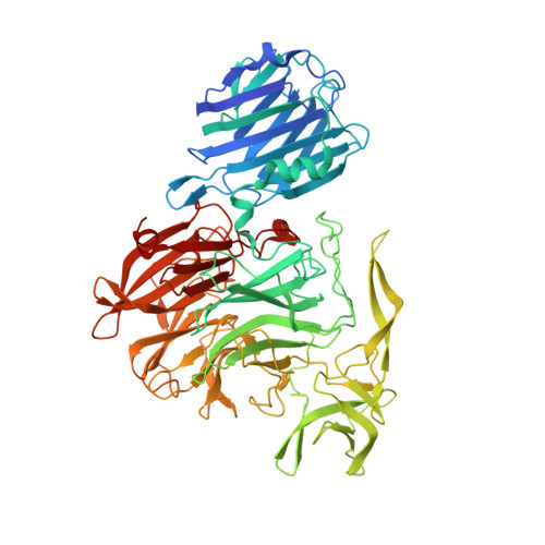
Streptococcus pneumoniae NanC: STRUCTURAL INSIGHTS INTO THE SPECIFICITY AND MECHANISM OF A SIALIDASE THAT PRODUCES A SIALIDASE INHIBITOR.

Owen, C.D.Lukacik, P.Potter, J.A.Sleator, O.Taylor, G.L.Walsh, M.A.

(2015) J Biological Chem 290: 27736-27748

  • DOI: https://doi.org/10.1074/jbc.M115.673632
  • Primary Citation Related Structures: 
    4YW1, 4YW2, 4YW3, 4YW5, 4YZ1, 4YZ2, 4YZ4, 4YZ5, 5F9T

  • PubMed Abstract: 

    Streptococcus pneumoniae is an important human pathogen that causes a range of disease states. Sialidases are important bacterial virulence factors. There are three pneumococcal sialidases: NanA, NanB, and NanC. NanC is an unusual sialidase in that its primary reaction product is 2-deoxy-2,3-didehydro-N-acetylneuraminic acid (Neu5Ac2en, also known as DANA), a nonspecific hydrolytic sialidase inhibitor. The production of Neu5Ac2en from α2-3-linked sialosides by the catalytic domain is confirmed within a crystal structure. A covalent complex with 3-fluoro-β-N-acetylneuraminic acid is also presented, suggesting a common mechanism with other sialidases up to the final step of product formation. A conformation change in an active site hydrophobic loop on ligand binding constricts the entrance to the active site. In addition, the distance between the catalytic acid/base (Asp-315) and the ligand anomeric carbon is unusually short. These features facilitate a novel sialidase reaction in which the final step of product formation is direct abstraction of the C3 proton by the active site aspartic acid, forming Neu5Ac2en. NanC also possesses a carbohydrate-binding module, which is shown to bind α2-3- and α2-6-linked sialosides, as well as N-acetylneuraminic acid, which is captured in the crystal structure following hydration of Neu5Ac2en by NanC. Overall, the pneumococcal sialidases show remarkable mechanistic diversity while maintaining a common structural scaffold.


  • Organizational Affiliation
    • From the Biomedical Sciences Research Complex, University of St. Andrews, St. Andrews, Fife KY16 9ST, United Kingdom.

Macromolecule Content 

  • Total Structure Weight: 153.76 kDa 
  • Atom Count: 11,166 
  • Modeled Residue Count: 1,311 
  • Deposited Residue Count: 1,354 
  • Unique protein chains: 1

Macromolecules

Find similar proteins by:|  3D Structure
Entity ID: 1
MoleculeChains  Sequence LengthOrganismDetailsImage
Sialidase, NanC
A, B
677Streptococcus pneumoniaeMutation(s): 0 
Gene Names: SP_1326
EC: 3.2.1.18
UniProt
Find proteins for A0A0H2UQE4 (Streptococcus pneumoniae serotype 4 (strain ATCC BAA-334 / TIGR4))
Explore A0A0H2UQE4 
Go to UniProtKB:  A0A0H2UQE4
Entity Groups
Sequence Clusters30% Identity50% Identity70% Identity90% Identity95% Identity100% Identity
UniProt GroupA0A0H2UQE4
Sequence Annotations
Expand
Reference Sequence

Small Molecules

Ligands 2 Unique
IDChains Name / Formula / InChI Key2D Diagram3D Interactions
SIA

Query on SIA


Download:Ideal Coordinates CCD File
C [auth A],
J [auth B]
N-acetyl-alpha-neuraminic acid
C11 H19 N O9
SQVRNKJHWKZAKO-YRMXFSIDSA-N
SO4

Query on SO4


Download:Ideal Coordinates CCD File
D [auth A]
E [auth A]
F [auth A]
G [auth A]
H [auth A]
D [auth A],
E [auth A],
F [auth A],
G [auth A],
H [auth A],
I [auth A],
K [auth B],
L [auth B],
M [auth B],
N [auth B],
O [auth B],
P [auth B]
SULFATE ION
O4 S
QAOWNCQODCNURD-UHFFFAOYSA-L

Experimental Data & Validation

Experimental Data

  • Method: X-RAY DIFFRACTION
  • Resolution: 2.15 Å
  • R-Value Free:  0.239 (Depositor), 0.241 (DCC) 
  • R-Value Work:  0.203 (Depositor), 0.210 (DCC) 
  • R-Value Observed: 0.205 (Depositor) 
Space Group: P 21 21 21
Unit Cell:
Length ( Å )Angle ( ˚ )
a = 73.38α = 90
b = 136.09β = 90
c = 150.44γ = 90
Software Package:
Software NamePurpose
REFMACrefinement
PDB_EXTRACTdata extraction
xia2data reduction
xia2data scaling

Structure Validation

View Full Validation Report


Ligand Structure Quality Assessment 


Entry History 

& Funding Information

Deposition Data


Funding OrganizationLocationGrant Number
Medical Research Council (United Kingdom)United Kingdom--

Revision History  (Full details and data files)

  • Version 1.0: 2015-09-23
    Type: Initial release
  • Version 1.1: 2015-11-25
    Changes: Database references
  • Version 1.2: 2017-09-13
    Changes: Author supporting evidence
  • Version 1.3: 2020-07-29
    Type: Remediation
    Reason: Carbohydrate remediation
    Changes: Data collection, Derived calculations, Structure summary
  • Version 1.4: 2024-05-08
    Changes: Data collection, Database references, Refinement description, Structure summary