4YXW

Bovine heart mitochondrial F1-ATPase inhibited by AMP-PNP and ADP in the presence of thiophosphate.


Experimental Data Snapshot

  • Method: X-RAY DIFFRACTION
  • Resolution: 3.10 Å
  • R-Value Free: 0.273 
  • R-Value Work: 0.226 
  • R-Value Observed: 0.229 

Starting Model: experimental
View more details

wwPDB Validation   3D Report Full Report


Ligand Structure Quality Assessment 


This is version 1.3 of the entry. See complete history


Literature

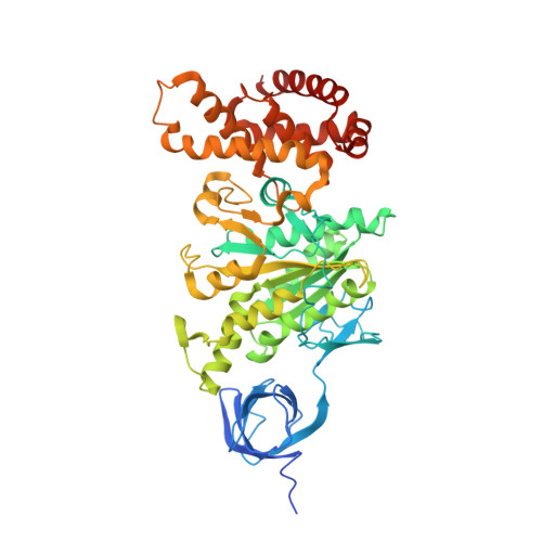
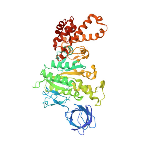
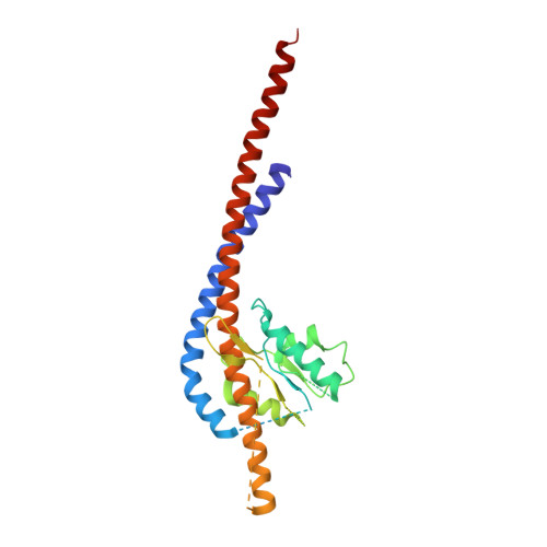
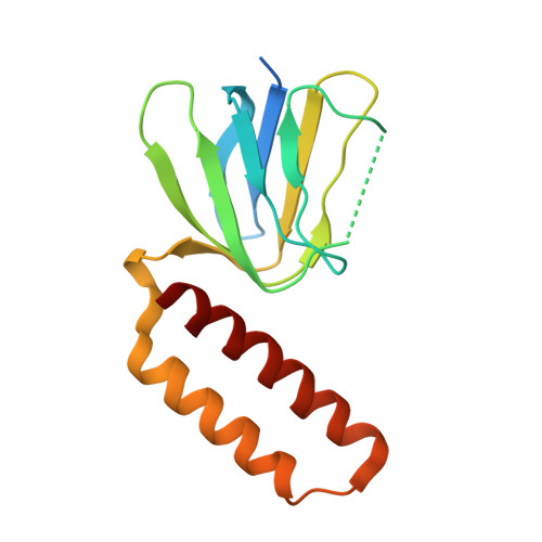
How release of phosphate from mammalian F1-ATPase generates a rotary substep.

Bason, J.V.Montgomery, M.G.Leslie, A.G.Walker, J.E.

(2015) Proc Natl Acad Sci U S A 112: 6009-6014

  • DOI: https://doi.org/10.1073/pnas.1506465112
  • Primary Citation of Related Structures:  
    4YXW, 4Z1M

  • PubMed Abstract: 

    The rotation of the central stalk of F1-ATPase is driven by energy derived from the sequential binding of an ATP molecule to its three catalytic sites and the release of the products of hydrolysis. In human F1-ATPase, each 360° rotation consists of three 120° steps composed of substeps of about 65°, 25°, and 30°, with intervening ATP binding, phosphate release, and catalytic dwells, respectively. The F1-ATPase inhibitor protein, IF1, halts the rotary cycle at the catalytic dwell. The human and bovine enzymes are essentially identical, and the structure of bovine F1-ATPase inhibited by IF1 represents the catalytic dwell state. Another structure, described here, of bovine F1-ATPase inhibited by an ATP analog and the phosphate analog, thiophosphate, represents the phosphate binding dwell. Thiophosphate is bound to a site in the α(E)β(E)-catalytic interface, whereas in F1-ATPase inhibited with IF1, the equivalent site is changed subtly and the enzyme is incapable of binding thiophosphate. These two structures provide a molecular mechanism of how phosphate release generates a rotary substep as follows. In the active enzyme, phosphate release from the β(E)-subunit is accompanied by a rearrangement of the structure of its binding site that prevents released phosphate from rebinding. The associated extrusion of a loop in the β(E)-subunit disrupts interactions in the α(E)β(E-)catalytic interface and opens it to its fullest extent. Other rearrangements disrupt interactions between the γ-subunit and the C-terminal domain of the α(E)-subunit. To restore most of these interactions, and to make compensatory new ones, the γ-subunit rotates through 25°-30°.


  • Organizational Affiliation

    Medical Research Council Mitochondrial Biology Unit, Cambridge Biomedical Campus, Hills Road, Cambridge CB2 0XY, United Kingdom; and.


Macromolecules
Find similar proteins by:  (by identity cutoff)  |  3D Structure
Entity ID: 1
MoleculeChains Sequence LengthOrganismDetailsImage
ATP synthase subunit alpha, mitochondrial
A, B, C
510Bos taurusMutation(s): 2 
Membrane Entity: Yes 
UniProt
Find proteins for P19483 (Bos taurus)
Explore P19483 
Go to UniProtKB:  P19483
Entity Groups  
Sequence Clusters30% Identity50% Identity70% Identity90% Identity95% Identity100% Identity
UniProt GroupP19483
Sequence Annotations
Expand
  • Reference Sequence
Find similar proteins by:  (by identity cutoff)  |  3D Structure
Entity ID: 2
MoleculeChains Sequence LengthOrganismDetailsImage
ATP synthase subunit beta, mitochondrial
D, E, F
482Bos taurusMutation(s): 0 
EC: 3.6.3.14
Membrane Entity: Yes 
UniProt
Find proteins for P00829 (Bos taurus)
Explore P00829 
Go to UniProtKB:  P00829
Entity Groups  
Sequence Clusters30% Identity50% Identity70% Identity90% Identity95% Identity100% Identity
UniProt GroupP00829
Sequence Annotations
Expand
  • Reference Sequence
Find similar proteins by:  (by identity cutoff)  |  3D Structure
Entity ID: 3
MoleculeChains Sequence LengthOrganismDetailsImage
ATP synthase subunit gamma, mitochondrial273Bos taurusMutation(s): 0 
Membrane Entity: Yes 
UniProt
Find proteins for P05631 (Bos taurus)
Explore P05631 
Go to UniProtKB:  P05631
Entity Groups  
Sequence Clusters30% Identity50% Identity70% Identity90% Identity95% Identity100% Identity
UniProt GroupP05631
Sequence Annotations
Expand
  • Reference Sequence
Find similar proteins by:  (by identity cutoff)  |  3D Structure
Entity ID: 4
MoleculeChains Sequence LengthOrganismDetailsImage
ATP synthase subunit delta, mitochondrial146Bos taurusMutation(s): 0 
Membrane Entity: Yes 
UniProt
Find proteins for P05630 (Bos taurus)
Explore P05630 
Go to UniProtKB:  P05630
Entity Groups  
Sequence Clusters30% Identity50% Identity70% Identity90% Identity95% Identity100% Identity
UniProt GroupP05630
Sequence Annotations
Expand
  • Reference Sequence
Find similar proteins by:  (by identity cutoff)  |  3D Structure
Entity ID: 5
MoleculeChains Sequence LengthOrganismDetailsImage
ATP synthase subunit epsilon, mitochondrial50Bos taurusMutation(s): 0 
Membrane Entity: Yes 
UniProt
Find proteins for P05632 (Bos taurus)
Explore P05632 
Go to UniProtKB:  P05632
Entity Groups  
Sequence Clusters30% Identity50% Identity70% Identity90% Identity95% Identity100% Identity
UniProt GroupP05632
Sequence Annotations
Expand
  • Reference Sequence
Small Molecules
Ligands 5 Unique
IDChains Name / Formula / InChI Key2D Diagram3D Interactions
ANP
Query on ANP

Download Ideal Coordinates CCD File 
J [auth A],
L [auth B],
N [auth C],
P [auth D],
T [auth F]
PHOSPHOAMINOPHOSPHONIC ACID-ADENYLATE ESTER
C10 H17 N6 O12 P3
PVKSNHVPLWYQGJ-KQYNXXCUSA-N
TS6
Query on TS6

Download Ideal Coordinates CCD File 
S [auth E]Monothiophosphate
H3 O3 P S
RYYWUUFWQRZTIU-UHFFFAOYSA-N
CL
Query on CL

Download Ideal Coordinates CCD File 
R [auth D]CHLORIDE ION
Cl
VEXZGXHMUGYJMC-UHFFFAOYSA-M
MG
Query on MG

Download Ideal Coordinates CCD File 
K [auth A],
M [auth B],
O [auth C],
Q [auth D],
U [auth F]
MAGNESIUM ION
Mg
JLVVSXFLKOJNIY-UHFFFAOYSA-N
NA
Query on NA

Download Ideal Coordinates CCD File 
V [auth F]SODIUM ION
Na
FKNQFGJONOIPTF-UHFFFAOYSA-N
Experimental Data & Validation

Experimental Data

  • Method: X-RAY DIFFRACTION
  • Resolution: 3.10 Å
  • R-Value Free: 0.273 
  • R-Value Work: 0.226 
  • R-Value Observed: 0.229 
  • Space Group: P 21 21 21
Unit Cell:
Length ( Å )Angle ( ˚ )
a = 105.353α = 90
b = 123.052β = 90
c = 261.276γ = 90
Software Package:
Software NamePurpose
MOSFLMdata reduction
Aimlessdata scaling
PHASERphasing
REFMACrefinement
Cootmodel building
PDB_EXTRACTdata extraction

Structure Validation

View Full Validation Report


Ligand Structure Quality Assessment 


Entry History 

Deposition Data

Revision History  (Full details and data files)

  • Version 1.0: 2015-05-06
    Type: Initial release
  • Version 1.1: 2015-05-13
    Changes: Database references
  • Version 1.2: 2015-05-20
    Changes: Database references
  • Version 1.3: 2024-01-10
    Changes: Data collection, Database references, Derived calculations, Refinement description