4YVV

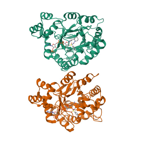
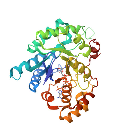
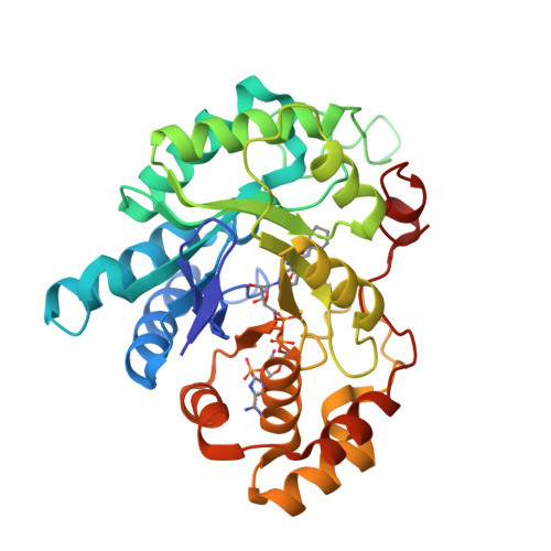
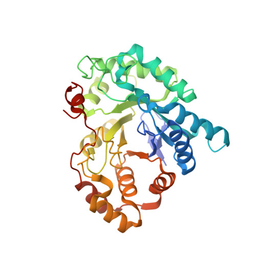
Crystal structure of AKR1C3 complexed with glibenclamide


Experimental Data Snapshot

  • Method: X-RAY DIFFRACTION
  • Resolution: 2.30 Å
  • R-Value Free: 0.253 
  • R-Value Work: 0.198 
  • R-Value Observed: 0.201 

Starting Model: experimental
View more details

wwPDB Validation   3D Report Full Report


Ligand Structure Quality Assessment 


This is version 1.2 of the entry. See complete history


Literature

In vitro inhibition of AKR1Cs by sulphonylureas and the structural basis

Zhao, Y.Zheng, X.Zhang, H.Zhai, J.Zhang, L.Li, C.Zeng, K.Chen, Y.Li, Q.Hu, X.

(2015) Chem Biol Interact 240: 310-315

  • DOI: https://doi.org/10.1016/j.cbi.2015.09.006
  • Primary Citation of Related Structures:  
    4YVP, 4YVV, 4YVX, 4ZFC

  • PubMed Abstract: 

    Recent epidemiological studies show conflicting data for the first-line anti-diabetic sulphonylureas drugs in treating cancer progression in type II diabetes patients. How sulphonylureas promote or diminish tumor growth is not fully understood. Here, we report that seven sulphonylureas exhibit different in vitro inhibition towards AKR1Cs (AKR1C1, AKR1C2, AKR1C3), which are critical steroid hormone metabolism enzymes that are related to prostate cancer, breast cancer and endometrial diseases. Interactions of the sulphonylureas and AKR1Cs were analyzed by X-ray crystallography.


  • Organizational Affiliation

    School of Pharmaceutical Sciences & Centre for Cellular and Structural Biology of Sun Yat-sen University, Guangzhou 510006, China.


Macromolecules
Find similar proteins by:  (by identity cutoff)  |  3D Structure
Entity ID: 1
MoleculeChains Sequence LengthOrganismDetailsImage
Aldo-keto reductase family 1 member C3
A, B
323Homo sapiensMutation(s): 0 
Gene Names: AKR1C3
EC: 1 (PDB Primary Data), 1.1.1.357 (PDB Primary Data), 1.1.1.112 (PDB Primary Data), 1.1.1.188 (PDB Primary Data), 1.1.1.239 (PDB Primary Data), 1.1.1.64 (PDB Primary Data), 1.3.1.20 (PDB Primary Data)
UniProt & NIH Common Fund Data Resources
Find proteins for P42330 (Homo sapiens)
Go to UniProtKB:  P42330
PHAROS:  P42330
GTEx:  ENSG00000196139 
Sequence Annotations
Expand
  • Reference Sequence
Small Molecules
Ligands 2 Unique
IDChains Name / Formula / InChI Key2D Diagram3D Interactions
NAP
Query on NAP

Download Ideal Coordinates CCD File 
C [auth A],
E [auth B]
NADP NICOTINAMIDE-ADENINE-DINUCLEOTIDE PHOSPHATE
C21 H28 N7 O17 P3
XJLXINKUBYWONI-NNYOXOHSSA-N
GBM
Query on GBM

Download Ideal Coordinates CCD File 
D [auth A],
F [auth B]
5-chloro-N-(2-{4-[(cyclohexylcarbamoyl)sulfamoyl]phenyl}ethyl)-2-methoxybenzamide
C23 H28 Cl N3 O5 S
ZNNLBTZKUZBEKO-UHFFFAOYSA-N
Experimental Data & Validation

Experimental Data

  • Method: X-RAY DIFFRACTION
  • Resolution: 2.30 Å
  • R-Value Free: 0.253 
  • R-Value Work: 0.198 
  • R-Value Observed: 0.201 
  • Space Group: P 1
Unit Cell:
Length ( Å )Angle ( ˚ )
a = 47.103α = 74.49
b = 48.973β = 86.64
c = 83.367γ = 69.44
Software Package:
Software NamePurpose
REFMACrefinement
SCALAdata scaling
PDB_EXTRACTdata extraction

Structure Validation

View Full Validation Report


Ligand Structure Quality Assessment 


Entry History 

Deposition Data

Revision History  (Full details and data files)

  • Version 1.0: 2015-11-25
    Type: Initial release
  • Version 1.1: 2016-04-06
    Changes: Non-polymer description
  • Version 1.2: 2023-11-08
    Changes: Data collection, Database references, Derived calculations, Refinement description, Structure summary