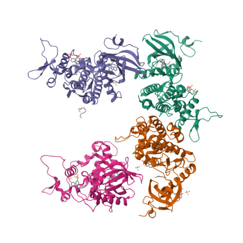
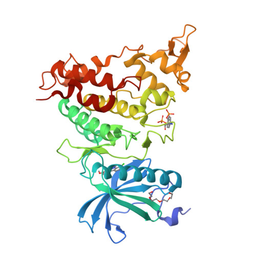
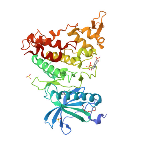
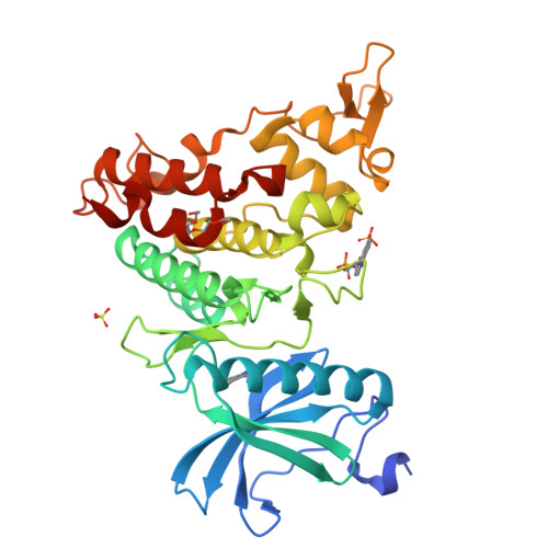
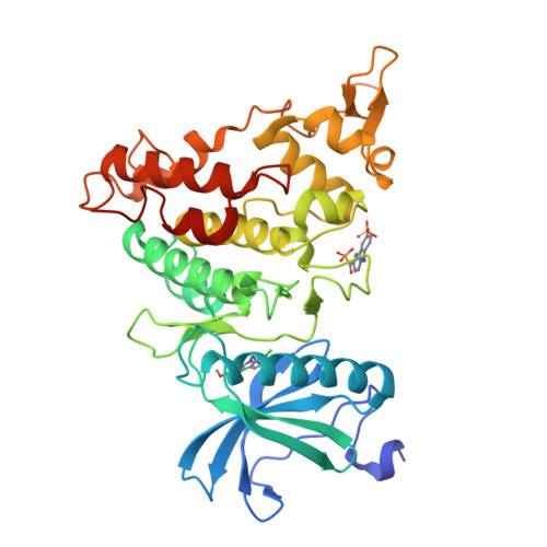
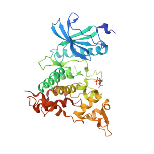
4YU2

Crystal structure of DYRK1A with harmine-derivatized AnnH-75 inhibitor


Experimental Data Snapshot

  • Method: X-RAY DIFFRACTION
  • Resolution: 2.90 Å
  • R-Value Free: 0.277 
  • R-Value Work: 0.243 
  • R-Value Observed: 0.245 

Starting Model: experimental
View more details

wwPDB Validation   3D Report Full Report


Ligand Structure Quality Assessment 


This is version 1.3 of the entry. See complete history


Literature

How to Separate Kinase Inhibition from Undesired Monoamine Oxidase A Inhibition-The Development of the DYRK1A Inhibitor AnnH75 from the Alkaloid Harmine.

Wurzlbauer, A.Ruben, K.Gurdal, E.Chaikuad, A.Knapp, S.Sippl, W.Becker, W.Bracher, F.

(2020) Molecules 25

  • DOI: https://doi.org/10.3390/molecules25245962
  • Primary Citation of Related Structures:  
    4YU2

  • PubMed Abstract: 

    The β-carboline alkaloid harmine is a potent DYRK1A inhibitor, but suffers from undesired potent inhibition of MAO-A, which strongly limits its application. We synthesized more than 60 analogues of harmine, either by direct modification of the alkaloid or by de novo synthesis of β-carboline and related scaffolds aimed at learning about structure-activity relationships for inhibition of both DYRK1A and MAO-A, with the ultimate goal of separating desired DYRK1A inhibition from undesired MAO-A inhibition. Based on evidence from published crystal structures of harmine bound to each of these enzymes, we performed systematic structure modifications of harmine yielding DYRK1A-selective inhibitors characterized by small polar substituents at N-9 (which preserve DYRK1A inhibition and eliminate MAO-A inhibition) and beneficial residues at C-1 (methyl or chlorine). The top compound AnnH75 remains a potent DYRK1A inhibitor, and it is devoid of MAO-A inhibition. Its binding mode to DYRK1A was elucidated by crystal structure analysis, and docking experiments provided additional insights for this attractive series of DYRK1A and MAO-A inhibitors.


  • Organizational Affiliation

    Department of Pharmacy-Center for Drug Research, Ludwig-Maximilians University, 81377 Munich, Germany.


Macromolecules
Find similar proteins by:  (by identity cutoff)  |  3D Structure
Entity ID: 1
MoleculeChains Sequence LengthOrganismDetailsImage
Dual specificity tyrosine-phosphorylation-regulated kinase 1A
A, B, C, D
361Homo sapiensMutation(s): 0 
Gene Names: DYRK1ADYRKMNBMNBH
EC: 2.7.12.1 (PDB Primary Data), 2.7.11.23 (UniProt)
UniProt & NIH Common Fund Data Resources
Find proteins for Q13627 (Homo sapiens)
Explore Q13627 
Go to UniProtKB:  Q13627
PHAROS:  Q13627
GTEx:  ENSG00000157540 
Entity Groups  
Sequence Clusters30% Identity50% Identity70% Identity90% Identity95% Identity100% Identity
UniProt GroupQ13627
Sequence Annotations
Expand
  • Reference Sequence
Small Molecules
Ligands 3 Unique
IDChains Name / Formula / InChI Key2D Diagram3D Interactions
4H5
Query on 4H5

Download Ideal Coordinates CCD File 
F [auth A],
H [auth B],
M [auth C],
O [auth D]
(1-chloro-7-methoxy-9H-beta-carbolin-9-yl)acetonitrile
C14 H10 Cl N3 O
BKIAHCJPMJSHSQ-UHFFFAOYSA-N
PG4
Query on PG4

Download Ideal Coordinates CCD File 
E [auth A],
G [auth B],
L [auth C]
TETRAETHYLENE GLYCOL
C8 H18 O5
UWHCKJMYHZGTIT-UHFFFAOYSA-N
SO4
Query on SO4

Download Ideal Coordinates CCD File 
I [auth B],
J [auth B],
K [auth B],
N [auth C]
SULFATE ION
O4 S
QAOWNCQODCNURD-UHFFFAOYSA-L
Modified Residues  1 Unique
IDChains TypeFormula2D DiagramParent
PTR
Query on PTR
A, B, C, D
L-PEPTIDE LINKINGC9 H12 N O6 PTYR
Binding Affinity Annotations 
IDSourceBinding Affinity
4H5 BindingDB:  4YU2 Kd: 6 (nM) from 1 assay(s)
IC50: min: 17, max: 181 (nM) from 3 assay(s)
Experimental Data & Validation

Experimental Data

  • Method: X-RAY DIFFRACTION
  • Resolution: 2.90 Å
  • R-Value Free: 0.277 
  • R-Value Work: 0.243 
  • R-Value Observed: 0.245 
  • Space Group: C 1 2 1
Unit Cell:
Length ( Å )Angle ( ˚ )
a = 265.567α = 90
b = 65.444β = 114.44
c = 138.648γ = 90
Software Package:
Software NamePurpose
REFMACrefinement
XDSdata reduction
SCALAdata scaling
PHASERphasing

Structure Validation

View Full Validation Report


Ligand Structure Quality Assessment 


Entry History 

Revision History  (Full details and data files)

  • Version 1.0: 2015-03-25
    Type: Initial release
  • Version 1.1: 2021-01-13
    Changes: Database references
  • Version 1.2: 2024-01-10
    Changes: Data collection, Database references, Refinement description
  • Version 1.3: 2024-11-20
    Changes: Structure summary