4YOW | pdb_00004yow

Crystal structure of a trimeric exonuclease PhoExo I from Pyrococcus horikoshii OT3 in complex with poly-dC


Experimental Data Snapshot

  • Method: X-RAY DIFFRACTION
  • Resolution: 2.50 Å
  • R-Value Free: 
    0.240 (Depositor), 0.240 (DCC) 
  • R-Value Work: 
    0.202 (Depositor), 0.204 (DCC) 
  • R-Value Observed: 
    0.204 (Depositor) 

wwPDB Validation 3D Report Full Report

Validation slider image for 4YOW

This is version 1.3 of the entry. See complete history

Literature

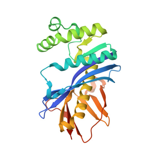
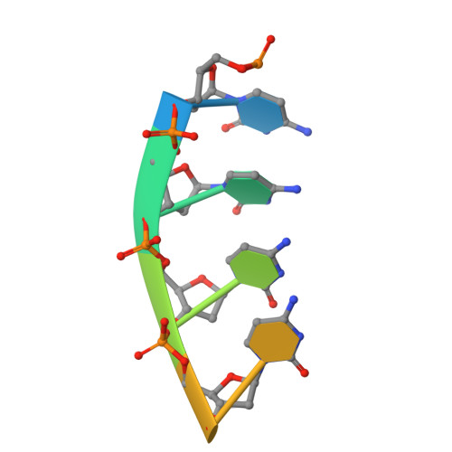
Structural basis for substrate recognition and processive cleavage mechanisms of the trimeric exonuclease PhoExo I

Miyazono, K.Ishino, S.Tsutsumi, K.Ito, T.Ishino, Y.Tanokura, M.

(2015) Nucleic Acids Res 43: 7122-7136

  • DOI: https://doi.org/10.1093/nar/gkv654
  • Primary Citation Related Structures: 
    4YOR, 4YOT, 4YOU, 4YOV, 4YOW, 4YOX, 4YOY

  • PubMed Abstract: 

    Nucleases play important roles in nucleic acid processes, such as replication, repair and recombination. Recently, we identified a novel single-strand specific 3'-5' exonuclease, PfuExo I, from the hyperthermophilic archaeon Pyrococcus furiosus, which may be involved in the Thermococcales-specific DNA repair system. PfuExo I forms a trimer and cleaves single-stranded DNA at every two nucleotides. Here, we report the structural basis for the cleavage mechanism of this novel exonuclease family. A structural analysis of PhoExo I, the homologous enzyme from P. horikoshii OT3, showed that PhoExo I utilizes an RNase H-like active site and possesses a 3'-OH recognition site ∼9 Å away from the active site, which enables cleavage at every two nucleotides. Analyses of the heterotrimeric and monomeric PhoExo I activities showed that trimerization is indispensable for its processive cleavage mechanism, but only one active site of the trimer is required.


  • Organizational Affiliation
    • Department of Applied Biological Chemistry, Graduate School of Agricultural and Life Sciences, The University of Tokyo, Tokyo, Japan.

Macromolecule Content 

  • Total Structure Weight: 84.6 kDa 
  • Atom Count: 5,666 
  • Modeled Residue Count: 693 
  • Deposited Residue Count: 720 
  • Unique protein chains: 1
  • Unique nucleic acid chains: 1

Macromolecules


Find similar proteins by:|  3D Structure
Entity ID: 1
MoleculeChains  Sequence LengthOrganismDetailsImage
3-5 exonuclease PhoExo I
A, C, E
233Pyrococcus horikoshiiMutation(s): 1 
UniProt
Find proteins for A0A060P168 (Pyrococcus horikoshii)
Explore A0A060P168 
Go to UniProtKB:  A0A060P168
Entity Groups
Sequence Clusters30% Identity50% Identity70% Identity90% Identity95% Identity100% Identity
UniProt GroupA0A060P168
Sequence Annotations
Expand
Reference Sequence
Find similar nucleic acids by:  Sequence
Entity ID: 2
MoleculeChains LengthOrganismImage
5'-D(*CP*CP*CP*CP*CP*CP*C)-3'
B, D, F
7synthetic construct
Sequence Annotations
Expand
Reference Sequence

Experimental Data & Validation

Experimental Data

  • Method: X-RAY DIFFRACTION
  • Resolution: 2.50 Å
  • R-Value Free:  0.240 (Depositor), 0.240 (DCC) 
  • R-Value Work:  0.202 (Depositor), 0.204 (DCC) 
  • R-Value Observed: 0.204 (Depositor) 
Space Group: P 21 21 21
Unit Cell:
Length ( Å )Angle ( ˚ )
a = 81.569α = 90
b = 100.265β = 90
c = 113.181γ = 90
Software Package:
Software NamePurpose
SCALAdata scaling
PHENIXrefinement
PDB_EXTRACTdata extraction
XDSdata reduction
MOLREPphasing

Structure Validation

View Full Validation Report


Entry History 

& Funding Information

Deposition Data


Funding OrganizationLocationGrant Number
Ministry of Education, Culture, Sports, Science, and TechnologyJapan--

Revision History  (Full details and data files)

  • Version 1.0: 2015-07-15
    Type: Initial release
  • Version 1.1: 2015-09-16
    Changes: Database references
  • Version 1.2: 2020-02-05
    Changes: Data collection, Database references, Derived calculations, Source and taxonomy
  • Version 1.3: 2024-03-20
    Changes: Data collection, Database references