4YOO

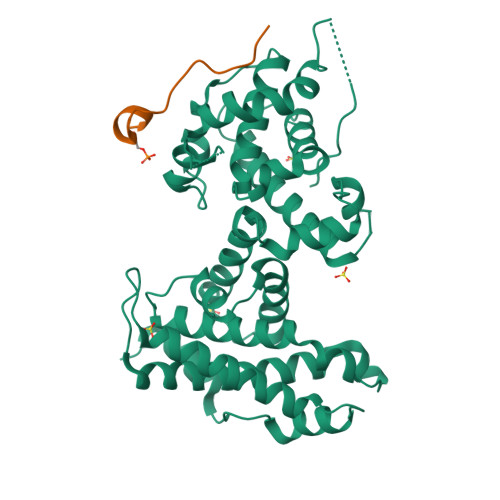
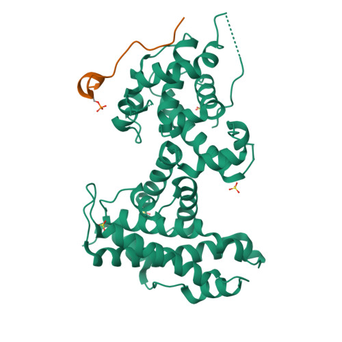
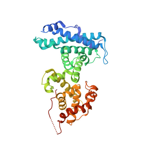
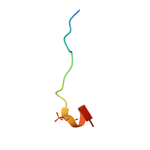
p107 pocket domain in complex with LIN52 P29A peptide


Experimental Data Snapshot

  • Method: X-RAY DIFFRACTION
  • Resolution: 2.40 Å
  • R-Value Free: 0.242 
  • R-Value Work: 0.195 
  • R-Value Observed: 0.200 

wwPDB Validation   3D Report Full Report


This is version 1.4 of the entry. See complete history


Literature

Structural mechanisms of DREAM complex assembly and regulation.

Guiley, K.Z.Liban, T.J.Felthousen, J.G.Ramanan, P.Litovchick, L.Rubin, S.M.

(2015) Genes Dev 29: 961-974

  • DOI: https://doi.org/10.1101/gad.257568.114
  • Primary Citation of Related Structures:  
    4YOO, 4YOS, 4YOZ

  • PubMed Abstract: 

    The DREAM complex represses cell cycle genes during quiescence through scaffolding MuvB proteins with E2F4/5 and the Rb tumor suppressor paralog p107 or p130. Upon cell cycle entry, MuvB dissociates from p107/p130 and recruits B-Myb and FoxM1 for up-regulating mitotic gene expression. To understand the biochemical mechanisms underpinning DREAM function and regulation, we investigated the structural basis for DREAM assembly. We identified a sequence in the MuvB component LIN52 that binds directly to the pocket domains of p107 and p130 when phosphorylated on the DYRK1A kinase site S28. A crystal structure of the LIN52-p107 complex reveals that LIN52 uses a suboptimal LxSxExL sequence together with the phosphate at nearby S28 to bind the LxCxE cleft of the pocket domain with high affinity. The structure explains the specificity for p107/p130 over Rb in the DREAM complex and how the complex is disrupted by viral oncoproteins. Based on insights from the structure, we addressed how DREAM is disassembled upon cell cycle entry. We found that p130 and B-Myb can both bind the core MuvB complex simultaneously but that cyclin-dependent kinase phosphorylation of p130 weakens its association. Together, our data inform a novel target interface for studying MuvB and p130 function and the design of inhibitors that prevent tumor escape in quiescence.


  • Organizational Affiliation

    Department of Chemistry and Biochemistry, University of California at Santa Cruz, Santa Cruz, California 95064, USA;


Macromolecules
Find similar proteins by:  (by identity cutoff)  |  3D Structure
Entity ID: 1
MoleculeChains Sequence LengthOrganismDetailsImage
Retinoblastoma-like protein 1,Retinoblastoma-like protein 1369Homo sapiensMutation(s): 0 
Gene Names: RBL1
UniProt & NIH Common Fund Data Resources
Find proteins for P28749 (Homo sapiens)
Explore P28749 
Go to UniProtKB:  P28749
PHAROS:  P28749
GTEx:  ENSG00000080839 
Entity Groups  
Sequence Clusters30% Identity50% Identity70% Identity90% Identity95% Identity100% Identity
UniProt GroupP28749
Sequence Annotations
Expand
  • Reference Sequence

Find similar proteins by:  Sequence   |   3D Structure  

Entity ID: 2
MoleculeChains Sequence LengthOrganismDetailsImage
LIN52 peptideB [auth X]20Homo sapiensMutation(s): 0 
UniProt & NIH Common Fund Data Resources
Find proteins for Q52LA3 (Homo sapiens)
Explore Q52LA3 
Go to UniProtKB:  Q52LA3
PHAROS:  Q52LA3
Entity Groups  
Sequence Clusters30% Identity50% Identity70% Identity90% Identity95% Identity100% Identity
UniProt GroupQ52LA3
Sequence Annotations
Expand
  • Reference Sequence
Experimental Data & Validation

Experimental Data

  • Method: X-RAY DIFFRACTION
  • Resolution: 2.40 Å
  • R-Value Free: 0.242 
  • R-Value Work: 0.195 
  • R-Value Observed: 0.200 
  • Space Group: C 2 2 21
Unit Cell:
Length ( Å )Angle ( ˚ )
a = 74.815α = 90
b = 100.53β = 90
c = 142.79γ = 90
Software Package:
Software NamePurpose
PHENIXrefinement
iMOSFLMdata reduction
SCALAdata scaling
Aimlessdata scaling

Structure Validation

View Full Validation Report


Entry History & Funding Information

Deposition Data


Funding OrganizationLocationGrant Number
National Institutes of Health/National Cancer Institute (NIH/NCI)United StatesR01CA132685

Revision History  (Full details and data files)

  • Version 1.0: 2015-05-13
    Type: Initial release
  • Version 1.1: 2015-05-20
    Changes: Database references
  • Version 1.2: 2017-09-20
    Changes: Author supporting evidence, Database references, Derived calculations, Refinement description, Source and taxonomy
  • Version 1.3: 2019-12-04
    Changes: Author supporting evidence
  • Version 1.4: 2024-10-30
    Changes: Data collection, Database references, Structure summary