4YO0 | pdb_00004yo0

Crystal structure of monoclonal anti-human podoplanin antibody NZ-1 with bound PA peptide


Experimental Data Snapshot

  • Method: X-RAY DIFFRACTION
  • Resolution: 1.56 Å
  • R-Value Free: 
    0.214 (Depositor), 0.215 (DCC) 
  • R-Value Work: 
    0.179 (Depositor), 0.180 (DCC) 
  • R-Value Observed: 
    0.181 (Depositor) 

Starting Model: experimental
View more details

wwPDB Validation 3D Report Full Report

Validation slider image for 4YO0

This is version 2.2 of the entry. See complete history

Literature

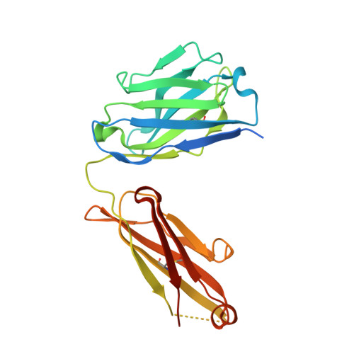
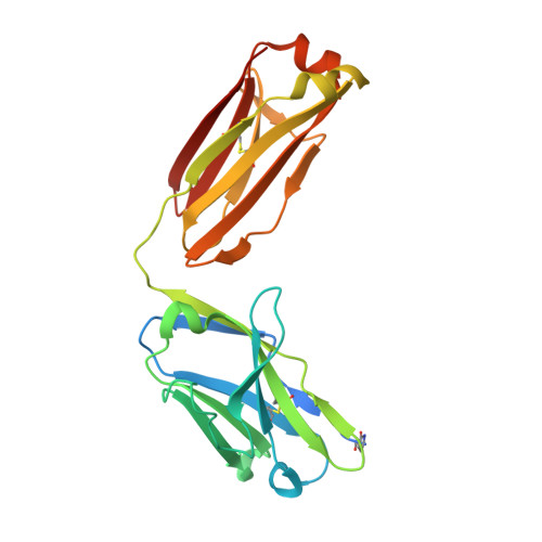
Tailored placement of a turn-forming PA tag into the structured domain of a protein to probe its conformational state

Fujii, Y.Matsunaga, Y.Arimori, T.Kitago, Y.Ogasawara, S.Kaneko, M.K.Kato, Y.Takagi, J.

(2016) J Cell Sci 129: 1512-1522

  • DOI: https://doi.org/10.1242/jcs.176685
  • Primary Citation Related Structures: 
    4YNY, 4YO0

  • PubMed Abstract: 

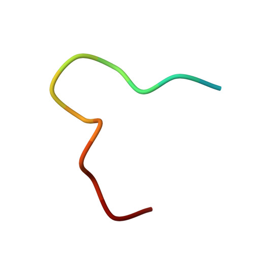
    Placement of a tag sequence is usually limited to either terminal end of the target protein, reducing the potential of epitope tags for various labeling applications. The PA tag is a dodecapeptide (GVAMPGAEDDVV) that is recognized by a high-affinity antibody NZ-1. We determined the crystal structure of the PA-tag-NZ-1 complex and found that NZ-1 recognizes a central segment of the PA tag peptide in a tight β-turn configuration, suggesting that it is compatible with the insertion into a loop. This possibility was tested and confirmed using multiple integrin subunits and semaphorin. More specifically, the PA tag can be inserted at multiple locations within the integrin αIIb subunit (encoded by ITGA2B) of the fibrinogen receptor αIIbβ3 integrin (of which the β3 subunit is encoded by ITGB3) without affecting the structural and functional integrity, while maintaining its high affinity for NZ-1. The large choice of the sites for 'epitope grafting' enabled the placement of the PA tag at a location whose accessibility is modulated during the biological action of the receptor. Thus, we succeeded in converting a general anti-tag antibody into a special anti-integrin antibody that can be classified as a ligand-induced binding site antibody.


  • Organizational Affiliation
    • Laboratory of Protein Synthesis and Expression, Institute for Protein Research, Osaka University, 3-2 Yamadaoka, Suita, Osaka 565-0871, Japan Department of Regional Innovation, Tohoku University Graduate School of Medicine, 2-1 Seiryo-machi, Aoba-ku, Sendai 980-8575, Japan.

Macromolecule Content 

  • Total Structure Weight: 104.86 kDa 
  • Atom Count: 7,078 
  • Modeled Residue Count: 850 
  • Deposited Residue Count: 970 
  • Unique protein chains: 3

Macromolecules

Find similar proteins by:|  3D Structure
Entity ID: 1
MoleculeChains  Sequence LengthOrganismDetailsImage
Heavy chain of antigen binding fragment, Fab
A, C
238Rattus norvegicusMutation(s): 0 
Find similar proteins by:|  3D Structure
Entity ID: 2
MoleculeChains  Sequence LengthOrganismDetailsImage
Light chain of antigen binding fragment, Fab
B, D
233Rattus norvegicusMutation(s): 0 
Find similar proteins by:  Sequence   |   3D Structure  
Entity ID: 3
MoleculeChains  Sequence LengthOrganismDetailsImage
PA14 peptide
E, F
14Homo sapiensMutation(s): 0 
UniProt & NIH Common Fund Data Resources
Find proteins for Q86YL7 (Homo sapiens)
Explore Q86YL7 
Go to UniProtKB:  Q86YL7
PHAROS:  Q86YL7
GTEx:  ENSG00000162493 
Entity Groups
Sequence Clusters30% Identity50% Identity70% Identity90% Identity95% Identity100% Identity
UniProt GroupQ86YL7
Sequence Annotations
Expand
Reference Sequence

Small Molecules

Ligands 1 Unique
IDChains Name / Formula / InChI Key2D Diagram3D Interactions
EDO

Query on EDO


Download:Ideal Coordinates CCD File
G [auth D]1,2-ETHANEDIOL
C2 H6 O2
LYCAIKOWRPUZTN-UHFFFAOYSA-N
Modified Residues  1 Unique
IDChains TypeFormula2D DiagramParent
PCA
Query on PCA
B, D
L-PEPTIDE LINKINGC5 H7 N O3GLN

Experimental Data & Validation

Experimental Data

  • Method: X-RAY DIFFRACTION
  • Resolution: 1.56 Å
  • R-Value Free:  0.214 (Depositor), 0.215 (DCC) 
  • R-Value Work:  0.179 (Depositor), 0.180 (DCC) 
  • R-Value Observed: 0.181 (Depositor) 
Space Group: P 1 21 1
Unit Cell:
Length ( Å )Angle ( ˚ )
a = 42.18α = 90
b = 80.01β = 96.61
c = 132.52γ = 90
Software Package:
Software NamePurpose
PHENIXrefinement
XDSdata reduction
XDSdata scaling
PHASERphasing

Structure Validation

View Full Validation Report


Entry History 

Deposition Data

Revision History  (Full details and data files)

  • Version 1.0: 2016-03-02
    Type: Initial release
  • Version 1.1: 2016-04-13
    Changes: Database references
  • Version 2.0: 2020-02-05
    Changes: Data collection, Derived calculations, Polymer sequence
  • Version 2.1: 2023-11-08
    Changes: Data collection, Database references, Refinement description
  • Version 2.2: 2024-11-20
    Changes: Structure summary