4YLI | pdb_00004yli

CL-K1 trimer


Experimental Data Snapshot

  • Method: X-RAY DIFFRACTION
  • Resolution: 2.45 Å
  • R-Value Free: 
    0.221 (Depositor), 0.226 (DCC) 
  • R-Value Work: 
    0.187 (Depositor), 0.193 (DCC) 
  • R-Value Observed: 
    0.189 (Depositor) 

Starting Model: experimental
View more details

wwPDB Validation 3D Report Full Report

Validation slider image for 4YLI

This is version 2.2 of the entry. See complete history

Literature

Molecular basis of sugar recognition by collectin-K1 and the effects of mutations associated with 3MC syndrome.

Girija, U.V.Furze, C.M.Gingras, A.R.Yoshizaki, T.Ohtani, K.Marshall, J.E.Wallis, A.K.Schwaeble, W.J.El-Mezgueldi, M.Mitchell, D.A.Moody, P.C.Wakamiya, N.Wallis, R.

(2015) BMC Biol 13: 27-27

  • DOI: https://doi.org/10.1186/s12915-015-0136-2
  • Primary Citation Related Structures: 
    4YLI, 4YMD

  • PubMed Abstract: 

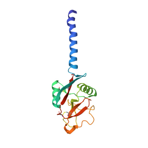
    Collectin-K1 (CL-K1, or CL-11) is a multifunctional Ca(2+)-dependent lectin with roles in innate immunity, apoptosis and embryogenesis. It binds to carbohydrates on pathogens to activate the lectin pathway of complement and together with its associated serine protease MASP-3 serves as a guidance cue for neural crest development. High serum levels are associated with disseminated intravascular coagulation, where spontaneous clotting can lead to multiple organ failure. Autosomal mutations in the CL-K1 or MASP-3 genes cause a developmental disorder called 3MC (Carnevale, Mingarelli, Malpuech and Michels) syndrome, characterised by facial, genital, renal and limb abnormalities. One of these mutations (Gly(204)Ser in the CL-K1 gene) is associated with undetectable levels of protein in the serum of affected individuals. In this study, we show that CL-K1 primarily targets a subset of high-mannose oligosaccharides present on both self- and non-self structures, and provide the structural basis for its ligand specificity. We also demonstrate that three disease-associated mutations prevent secretion of CL-K1 from mammalian cells, accounting for the protein deficiency observed in patients. Interestingly, none of the mutations prevent folding or oligomerization of recombinant fragments containing the mutations in vitro. Instead, they prevent Ca(2+) binding by the carbohydrate-recognition domains of CL-K1. We propose that failure to bind Ca(2+) during biosynthesis leads to structural defects that prevent secretion of CL-K1, thus providing a molecular explanation of the genetic disorder. We have established the sugar specificity of CL-K1 and demonstrated that it targets high-mannose oligosaccharides on self- and non-self structures via an extended binding site which recognises the terminal two mannose residues of the carbohydrate ligand. We have also shown that mutations associated with a rare developmental disorder called 3MC syndrome prevent the secretion of CL-K1, probably as a result of structural defects caused by disruption of Ca(2+) binding during biosynthesis.


  • Organizational Affiliation
    • Department of Infection, Immunity and Inflammation, University of Leicester, Leicester, LE1 9HN, UK. uvg1@leicester.ac.uk.

Macromolecule Content 

  • Total Structure Weight: 105.83 kDa 
  • Atom Count: 7,308 
  • Modeled Residue Count: 919 
  • Deposited Residue Count: 930 
  • Unique protein chains: 1

Macromolecules

Find similar proteins by:|  3D Structure
Entity ID: 1
MoleculeChains  Sequence LengthOrganismDetailsImage
Collectin-11
A, B, C, D, E
A, B, C, D, E, F
155Homo sapiensMutation(s): 0 
Gene Names: COLEC11UNQ596/PRO1182
UniProt & NIH Common Fund Data Resources
Find proteins for Q9BWP8 (Homo sapiens)
Explore Q9BWP8 
Go to UniProtKB:  Q9BWP8
PHAROS:  Q9BWP8
GTEx:  ENSG00000118004 
Entity Groups
Sequence Clusters30% Identity50% Identity70% Identity90% Identity95% Identity100% Identity
UniProt GroupQ9BWP8
Sequence Annotations
Expand
Reference Sequence

Small Molecules

Ligands 3 Unique
IDChains Name / Formula / InChI Key2D Diagram3D Interactions
GOL

Query on GOL


Download:Ideal Coordinates CCD File
AA [auth D]
BA [auth D]
FA [auth E]
GA [auth E]
K [auth A]
AA [auth D],
BA [auth D],
FA [auth E],
GA [auth E],
K [auth A],
KA [auth F],
L [auth A],
LA [auth F],
P [auth B],
Q [auth B],
R [auth B],
V [auth C],
W [auth C]
GLYCEROL
C3 H8 O3
PEDCQBHIVMGVHV-UHFFFAOYSA-N
CA

Query on CA


Download:Ideal Coordinates CCD File
CA [auth E]
DA [auth E]
EA [auth E]
G [auth A]
H [auth A]
CA [auth E],
DA [auth E],
EA [auth E],
G [auth A],
H [auth A],
HA [auth F],
I [auth A],
IA [auth F],
JA [auth F],
M [auth B],
N [auth B],
O [auth B],
S [auth C],
T [auth C],
U [auth C],
X [auth D],
Y [auth D],
Z [auth D]
CALCIUM ION
Ca
BHPQYMZQTOCNFJ-UHFFFAOYSA-N
CL

Query on CL


Download:Ideal Coordinates CCD File
J [auth A]CHLORIDE ION
Cl
VEXZGXHMUGYJMC-UHFFFAOYSA-M

Experimental Data & Validation

Experimental Data

  • Method: X-RAY DIFFRACTION
  • Resolution: 2.45 Å
  • R-Value Free:  0.221 (Depositor), 0.226 (DCC) 
  • R-Value Work:  0.187 (Depositor), 0.193 (DCC) 
  • R-Value Observed: 0.189 (Depositor) 
Space Group: P 1 21 1
Unit Cell:
Length ( Å )Angle ( ˚ )
a = 72.99α = 90
b = 107.59β = 91.49
c = 77.82γ = 90
Software Package:
Software NamePurpose
MOSFLMdata reduction
PHENIXrefinement
PDB_EXTRACTdata extraction
Aimlessdata scaling
PHASERphasing

Structure Validation

View Full Validation Report


Entry History 

& Funding Information

Deposition Data


Funding OrganizationLocationGrant Number
Medical Research Council (United Kingdom)United KingdomG1000191/1

Revision History  (Full details and data files)

  • Version 1.0: 2015-04-08
    Type: Initial release
  • Version 1.1: 2015-05-13
    Changes: Database references
  • Version 2.0: 2017-08-30
    Changes: Advisory, Atomic model, Author supporting evidence, Derived calculations
  • Version 2.1: 2024-01-10
    Changes: Data collection, Database references, Derived calculations, Refinement description
  • Version 2.2: 2024-11-06
    Changes: Structure summary