4YK1 | pdb_00004yk1

Crystal Structure of the BID Domain of Bep6 from Bartonella rochalimae


Experimental Data Snapshot

  • Method: X-RAY DIFFRACTION
  • Resolution: 2.10 Å
  • R-Value Free: 
    0.203 (Depositor), 0.203 (DCC) 
  • R-Value Work: 
    0.177 (Depositor), 0.178 (DCC) 
  • R-Value Observed: 
    0.178 (Depositor) 

wwPDB Validation 3D Report Full Report

Validation slider image for 4YK1

This is version 1.2 of the entry. See complete history

Literature

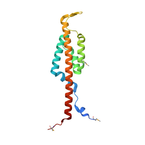
The BID Domain of Type IV Secretion Substrates Forms a Conserved Four-Helix Bundle Topped with a Hook.

Stanger, F.V.de Beer, T.A.Dranow, D.M.Schirmer, T.Phan, I.Dehio, C.

(2017) Structure 25: 203-211

  • DOI: https://doi.org/10.1016/j.str.2016.10.010
  • Primary Citation Related Structures: 
    4YK1, 4YK2, 4YK3

  • PubMed Abstract: 

    The BID (Bep intracellular delivery) domain functions as secretion signal in a subfamily of protein substrates of bacterial type IV secretion (T4S) systems. It mediates transfer of (1) relaxases and the attached DNA during bacterial conjugation, and (2) numerous Bartonella effector proteins (Beps) during protein transfer into host cells infected by pathogenic Bartonella species. Furthermore, BID domains of Beps have often evolved secondary effector functions within host cells. Here, we provide crystal structures for three representative BID domains and describe a novel conserved fold characterized by a compact, antiparallel four-helix bundle topped with a hook. The conserved hydrophobic core provides a rigid scaffold to a surface that, despite a few conserved exposed residues and similarities in charge distribution, displays significant variability. We propose that the genuine function of BID domains as T4S signal may primarily depend on their rigid structure, while the plasticity of their surface may facilitate adaptation to secondary effector functions.


  • Organizational Affiliation
    • Focal Area Infection Biology, Biozentrum University of Basel, 4056 Basel, Switzerland; Focal Area Structural Biology and Biophysics, Biozentrum University of Basel, 4056 Basel, Switzerland.

Macromolecule Content 

  • Total Structure Weight: 17.19 kDa 
  • Atom Count: 1,174 
  • Modeled Residue Count: 137 
  • Deposited Residue Count: 146 
  • Unique protein chains: 1

Macromolecules

Find similar proteins by:|  3D Structure
Entity ID: 1
MoleculeChains  Sequence LengthOrganismDetailsImage
Bartonella effector protein (Bep) substrate of VirB T4SS146Bartonella rochalimae ATCC BAA-1498Mutation(s): 0 
Gene Names: BARRO_50054O99_01278
EC: 2.7.7.108
UniProt
Find proteins for E6YLF3 (Bartonella rochalimae ATCC BAA-1498)
Explore E6YLF3 
Go to UniProtKB:  E6YLF3
Entity Groups
Sequence Clusters30% Identity50% Identity70% Identity90% Identity95% Identity100% Identity
UniProt GroupE6YLF3
Sequence Annotations
Expand
Reference Sequence

Small Molecules

Modified Residues  1 Unique
IDChains TypeFormula2D DiagramParent
MSE
Query on MSE
A
L-PEPTIDE LINKINGC5 H11 N O2 SeMET

Experimental Data & Validation

Experimental Data

  • Method: X-RAY DIFFRACTION
  • Resolution: 2.10 Å
  • R-Value Free:  0.203 (Depositor), 0.203 (DCC) 
  • R-Value Work:  0.177 (Depositor), 0.178 (DCC) 
  • R-Value Observed: 0.178 (Depositor) 
Space Group: P 41 2 2
Unit Cell:
Length ( Å )Angle ( ˚ )
a = 79.25α = 90
b = 79.25β = 90
c = 85.97γ = 90
Software Package:
Software NamePurpose
XSCALEdata scaling
PHASERphasing
PHENIXrefinement
PDB_EXTRACTdata extraction
XDSdata reduction
ARPmodel building

Structure Validation

View Full Validation Report


Entry History 

Deposition Data

Revision History  (Full details and data files)

  • Version 1.0: 2016-03-09
    Type: Initial release
  • Version 1.1: 2017-01-11
    Changes: Database references
  • Version 1.2: 2024-10-23
    Changes: Data collection, Database references, Structure summary