4YHA | pdb_00004yha

Crystal structure of the complex of Helicobacter pylori alpha-Carbonic Anhydrase with methazolamide


Experimental Data Snapshot

  • Method: X-RAY DIFFRACTION
  • Resolution: 2.20 Å
  • R-Value Free: 
    0.245 (Depositor), 0.240 (DCC) 
  • R-Value Work: 
    0.208 (Depositor), 0.206 (DCC) 
  • R-Value Observed: 
    0.212 (Depositor) 

Starting Model: experimental
View more details

wwPDB Validation 3D Report Full Report

Validation slider image for 4YHA

Ligand Structure Quality Assessment 


This is version 2.2 of the entry. See complete history

Literature

Structural Basis for the Inhibition of Helicobacter pylori alpha-Carbonic Anhydrase by Sulfonamides.

Modakh, J.K.Liu, Y.C.Machuca, M.A.Supuran, C.T.Roujeinikova, A.

(2015) PLoS One 10: e0127149-e0127149

  • DOI: https://doi.org/10.1371/journal.pone.0127149
  • Primary Citation Related Structures: 
    4YGF, 4YHA

  • PubMed Abstract: 

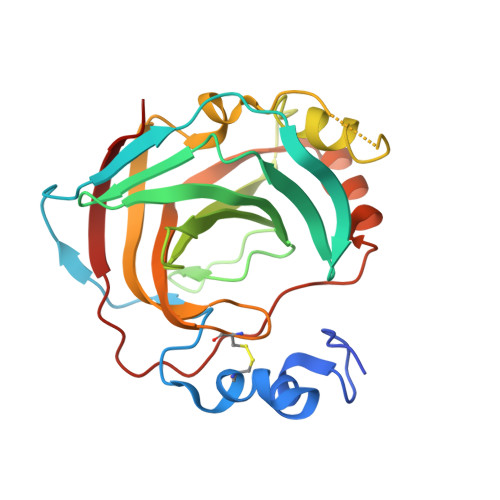
    Periplasmic α-carbonic anhydrase of Helicobacter pylori (HpαCA), an oncogenic bacterium in the human stomach, is essential for its acclimation to low pH. It catalyses the conversion of carbon dioxide to bicarbonate using Zn(II) as the cofactor. In H. pylori, Neisseria spp., Brucella suis and Streptococcus pneumoniae this enzyme is the target for sulfonamide antibacterial agents. We present structural analysis correlated with inhibition data, on the complexes of HpαCA with two pharmacological inhibitors of human carbonic anhydrases, acetazolamide and methazolamide. This analysis reveals that two sulfonamide oxygen atoms of the inhibitors are positioned proximal to the putative location of the oxygens of the CO2 substrate in the Michaelis complex, whilst the zinc-coordinating sulfonamide nitrogen occupies the position of the catalytic water molecule. The structures are consistent with acetazolamide acting as site-directed, nanomolar inhibitors of the enzyme by mimicking its reaction transition state. Additionally, inhibitor binding provides insights into the channel for substrate entry and product exit. This analysis has implications for the structure-based design of inhibitors of bacterial carbonic anhydrases.


  • Organizational Affiliation
    • Department of Microbiology, Faculty of Biomedical and Psychological Sciences, Monash University, Clayton, Victoria, Australia.

Macromolecule Content 

  • Total Structure Weight: 219.01 kDa 
  • Atom Count: 15,354 
  • Modeled Residue Count: 1,782 
  • Deposited Residue Count: 1,872 
  • Unique protein chains: 1

Macromolecules

Find similar proteins by:|  3D Structure
Entity ID: 1
MoleculeChains  Sequence LengthOrganismDetailsImage
Alpha-carbonic anhydrase
A, B, C, D, E
A, B, C, D, E, F, G, H
234Helicobacter pylori 26695Mutation(s): 0 
Gene Names: C694_06140
EC: 4.2.1.1
UniProt
Find proteins for A0A0M3KL20 (Helicobacter pylori (strain ATCC 700392 / 26695))
Explore A0A0M3KL20 
Go to UniProtKB:  A0A0M3KL20
Entity Groups
Sequence Clusters30% Identity50% Identity70% Identity90% Identity95% Identity100% Identity
UniProt GroupA0A0M3KL20
Sequence Annotations
Expand
Reference Sequence

Small Molecules

Ligands 4 Unique
IDChains Name / Formula / InChI Key2D Diagram3D Interactions
MZM

Query on MZM


Download:Ideal Coordinates CCD File
AA [auth F]
DA [auth G]
HA [auth H]
K [auth A]
N [auth B]
AA [auth F],
DA [auth G],
HA [auth H],
K [auth A],
N [auth B],
Q [auth C],
T [auth D],
X [auth E]
N-(3-methyl-5-sulfamoyl-1,3,4-thiadiazol-2(3H)-ylidene)acetamide
C5 H8 N4 O3 S2
FLOSMHQXBMRNHR-DAXSKMNVSA-N
GOL

Query on GOL


Download:Ideal Coordinates CCD File
EA [auth G],
R [auth C],
U [auth D],
Y [auth E]
GLYCEROL
C3 H8 O3
PEDCQBHIVMGVHV-UHFFFAOYSA-N
ZN

Query on ZN


Download:Ideal Coordinates CCD File
BA [auth G]
FA [auth H]
I [auth A]
L [auth B]
O [auth C]
BA [auth G],
FA [auth H],
I [auth A],
L [auth B],
O [auth C],
S [auth D],
V [auth E],
Z [auth F]
ZINC ION
Zn
PTFCDOFLOPIGGS-UHFFFAOYSA-N
CL

Query on CL


Download:Ideal Coordinates CCD File
CA [auth G]
GA [auth H]
J [auth A]
M [auth B]
P [auth C]
CA [auth G],
GA [auth H],
J [auth A],
M [auth B],
P [auth C],
W [auth E]
CHLORIDE ION
Cl
VEXZGXHMUGYJMC-UHFFFAOYSA-M
Binding Affinity Annotations 
IDSourceBinding Affinity
MZM BindingDB:  4YHA Ki: 225 (nM) from 1 assay(s)
CL BindingDB:  4YHA Ki: 2.70e+6 (nM) from 1 assay(s)

Experimental Data & Validation

Experimental Data

  • Method: X-RAY DIFFRACTION
  • Resolution: 2.20 Å
  • R-Value Free:  0.245 (Depositor), 0.240 (DCC) 
  • R-Value Work:  0.208 (Depositor), 0.206 (DCC) 
  • R-Value Observed: 0.212 (Depositor) 
Space Group: P 1 21 1
Unit Cell:
Length ( Å )Angle ( ˚ )
a = 42.45α = 90
b = 135.65β = 90.05
c = 166.58γ = 90
Software Package:
Software NamePurpose
PHENIXrefinement
SCALAdata scaling
PDB_EXTRACTdata extraction
MOSFLMdata reduction
Cootmodel building
iMOSFLMdata reduction
PHASERphasing

Structure Validation

View Full Validation Report


Ligand Structure Quality Assessment 


Entry History 

Deposition Data

Revision History  (Full details and data files)

  • Version 1.0: 2015-07-01
    Type: Initial release
  • Version 1.1: 2015-12-16
    Changes: Structure summary
  • Version 2.0: 2017-11-22
    Changes: Data collection, Derived calculations, Non-polymer description, Refinement description, Structure summary
  • Version 2.1: 2023-09-27
    Changes: Data collection, Database references, Derived calculations, Refinement description
  • Version 2.2: 2024-11-20
    Changes: Structure summary