4YFY | pdb_00004yfy

X-ray structure of the Viof N-formyltransferase from Providencia alcalifaciens O30 in complex with THF and TDP-Qui4N


Experimental Data Snapshot

  • Method: X-RAY DIFFRACTION
  • Resolution: 1.90 Å
  • R-Value Free: 
    0.244 (Depositor), 0.249 (DCC) 
  • R-Value Work: 
    0.187 (Depositor), 0.196 (DCC) 
  • R-Value Observed: 
    0.189 (Depositor) 

Starting Model: experimental
View more details

wwPDB Validation 3D Report Full Report

Validation slider image for 4YFY

Ligand Structure Quality Assessment 


This is version 1.4 of the entry. See complete history

Literature

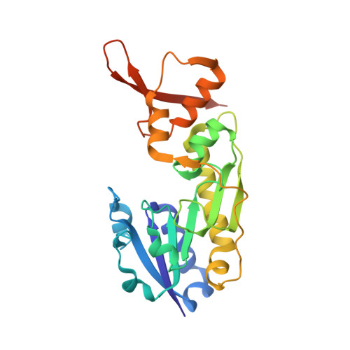
Molecular structure of an N-formyltransferase from Providencia alcalifaciens O30.

Genthe, N.A.Thoden, J.B.Benning, M.M.Holden, H.M.

(2015) Protein Sci 24: 976-986

  • DOI: https://doi.org/10.1002/pro.2675
  • Primary Citation Related Structures: 
    4YFV, 4YFY

  • PubMed Abstract: 

    The existence of N-formylated sugars in the O-antigens of Gram-negative bacteria has been known since the middle 1980s, but only recently have the biosynthetic pathways for their production been reported. In these pathways, glucose-1-phosphate is first activated by attachment to a dTMP moiety. This step is followed by a dehydration reaction and an amination. The last step in these pathways is catalyzed by N-formyltransferases that utilize N(10) -formyltetrahydrofolate as the carbon source. Here we describe the three-dimensional structure of one of these N-formyltransferases, namely VioF from Providencia alcalifaciens O30. Specifically, this enzyme catalyzes the conversion of dTDP-4-amino-4,6-dideoxyglucose (dTDP-Qui4N) to dTDP-4,6-dideoxy-4-formamido-d-glucose (dTDP-Qui4NFo). For this analysis, the structure of VioF was solved to 1.9 Å resolution in both its apoform and in complex with tetrahydrofolate and dTDP-Qui4N. The crystals used in the investigation belonged to the space group R32 and demonstrated reticular merohedral twinning. The overall catalytic core of the VioF subunit is characterized by a six stranded mixed β-sheet flanked on one side by three α-helices and on the other side by mostly random coil. This N-terminal domain is followed by an α-helix and a β-hairpin that form the subunit:subunit interface. The active site of the enzyme is shallow and solvent-exposed. Notably, the pyranosyl moiety of dTDP-Qui4N is positioned into the active site by only one hydrogen bond provided by Lys 77. Comparison of the VioF model to that of a previously determined N-formyltransferase suggests that substrate specificity is determined by interactions between the protein and the pyrophosphoryl group of the dTDP-sugar substrate.


  • Organizational Affiliation
    • Department of Biochemistry, University of Wisconsin, Madison, Wisconsin, 53706.

Macromolecule Content 

  • Total Structure Weight: 61.44 kDa 
  • Atom Count: 4,546 
  • Modeled Residue Count: 482 
  • Deposited Residue Count: 508 
  • Unique protein chains: 1

Macromolecules

Find similar proteins by:|  3D Structure
Entity ID: 1
MoleculeChains  Sequence LengthOrganismDetailsImage
VioF
A, B
254Providencia alcalifaciensMutation(s): 0 
Gene Names: vioF
UniProt
Find proteins for M9P0Q2 (Providencia alcalifaciens)
Explore M9P0Q2 
Go to UniProtKB:  M9P0Q2
Entity Groups
Sequence Clusters30% Identity50% Identity70% Identity90% Identity95% Identity100% Identity
UniProt GroupM9P0Q2
Sequence Annotations
Expand
Reference Sequence

Small Molecules

Ligands 4 Unique
IDChains Name / Formula / InChI Key2D Diagram3D Interactions
0FX

Query on 0FX


Download:Ideal Coordinates CCD File
C [auth A],
G [auth B]
dTDP-4-amino-4,6-dideoxyglucose
C16 H27 N3 O14 P2
UIVJXHWSIFBBCY-LPGAPTBISA-N
1YJ

Query on 1YJ


Download:Ideal Coordinates CCD File
D [auth A],
H [auth B]
N-[4-({[(6R)-2-amino-4-oxo-3,4,5,6,7,8-hexahydropteridin-6-yl]methyl}amino)benzoyl]-L-glutamic acid
C19 H23 N7 O6
MSTNYGQPCMXVAQ-NEPJUHHUSA-N
EDO

Query on EDO


Download:Ideal Coordinates CCD File
F [auth A]1,2-ETHANEDIOL
C2 H6 O2
LYCAIKOWRPUZTN-UHFFFAOYSA-N
CL

Query on CL


Download:Ideal Coordinates CCD File
E [auth A]CHLORIDE ION
Cl
VEXZGXHMUGYJMC-UHFFFAOYSA-M

Experimental Data & Validation

Experimental Data

  • Method: X-RAY DIFFRACTION
  • Resolution: 1.90 Å
  • R-Value Free:  0.244 (Depositor), 0.249 (DCC) 
  • R-Value Work:  0.187 (Depositor), 0.196 (DCC) 
  • R-Value Observed: 0.189 (Depositor) 
Space Group: H 3 2
Unit Cell:
Length ( Å )Angle ( ˚ )
a = 132α = 90
b = 132β = 90
c = 161.4γ = 120
Software Package:
Software NamePurpose
REFMACrefinement
SAINTdata reduction
SADABSdata scaling
PHASERphasing

Structure Validation

View Full Validation Report


Ligand Structure Quality Assessment 


Entry History 

Deposition Data

Revision History  (Full details and data files)

  • Version 1.0: 2015-03-11
    Type: Initial release
  • Version 1.1: 2015-03-25
    Changes: Structure summary
  • Version 1.2: 2015-04-01
    Changes: Database references
  • Version 1.3: 2015-06-03
    Changes: Database references
  • Version 1.4: 2023-09-27
    Changes: Data collection, Database references, Derived calculations, Other, Refinement description, Source and taxonomy