4YET | pdb_00004yet

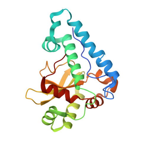
X-ray crystal structure of superoxide dismutase from Babesia bovis solved by Sulfur SAD


Experimental Data Snapshot

  • Method: X-RAY DIFFRACTION
  • Resolution: 1.75 Å
  • R-Value Free: 
    0.176 (Depositor), 0.179 (DCC) 
  • R-Value Work: 
    0.143 (Depositor), 0.146 (DCC) 
  • R-Value Observed: 
    0.145 (Depositor) 

wwPDB Validation 3D Report Full Report

Validation slider image for 4YET

This is version 1.5 of the entry. See complete history. 

Literature

Iron superoxide dismutases in eukaryotic pathogens: new insights from Apicomplexa and Trypanosoma structures.

Phan, I.Q., Davies, D.R., Moretti, N.S., Shanmugam, D., Cestari, I., Anupama, A., Fairman, J.W., Edwards, T.E., Stuart, K., Schenkman, S., Myler, P.J.

(2015) Acta Crystallogr F Struct Biol Commun 71: 615-621

  • DOI: https://doi.org/10.1107/S2053230X15004185
  • Primary Citation Related Structures: 
    4F2N, 4H3E, 4YET

  • PubMed Abstract: 

    Prior studies have highlighted the potential of superoxide dismutases as drug targets in eukaryotic pathogens. This report presents the structures of three iron-dependent superoxide dismutases (FeSODs) from Trypanosoma cruzi, Leishmania major and Babesia bovis. Comparison with existing structures from Plasmodium and other trypanosome isoforms shows a very conserved overall fold with subtle differences. In particular, structural data suggest that B. bovis FeSOD may display similar resistance to peroxynitrite-mediated inactivation via an intramolecular electron-transfer pathway as previously described in T. cruzi FeSOD isoform B, thus providing valuable information for structure-based drug design. Furthermore, lysine-acetylation results in T. cruzi indicate that acetylation occurs at a position close to that responsible for the regulation of acetylation-mediated activity in the human enzyme.


  • Organizational Affiliation: 
    • Seattle Structural Genomics Center for Infectious Disease (SSGCID), USA.

Macromolecule Content 

  • Total Structure Weight: 45.92 kDa 
  • Atom Count: 3,628 
  • Modeled Residue Count: 398 
  • Deposited Residue Count: 406 
  • Unique protein chains: 1

Macromolecules

Find similar proteins by:|  3D Structure
Entity ID: 1
MoleculeChains  Sequence LengthOrganismDetailsImage
Superoxide dismutase
A, B
203Babesia bovisMutation(s): 0 
Gene Names: Fe-SOD, BBOV_IV011480
EC: 1.15.1.1
UniProt
Find proteins for O15904 (Babesia bovis)
Explore O15904 
Go to UniProtKB:  O15904
Entity Groups
Sequence Clusters30% Identity50% Identity70% Identity90% Identity95% Identity100% Identity
UniProt GroupO15904
Sequence Annotations
Expand
Reference Sequence

Experimental Data & Validation

Experimental Data

  • Method: X-RAY DIFFRACTION
  • Resolution: 1.75 Å
  • R-Value Free:  0.176 (Depositor), 0.179 (DCC) 
  • R-Value Work:  0.143 (Depositor), 0.146 (DCC) 
  • R-Value Observed: 0.145 (Depositor) 
Space Group: P 21 21 21
Unit Cell:
Length ( Å )Angle ( ˚ )
a = 55.75α = 90
b = 69.09β = 90
c = 114.81γ = 90
Software Package:
Software NamePurpose
XSCALEdata scaling
PHENIXrefinement
PDB_EXTRACTdata extraction
PHASERphasing

Structure Validation

View Full Validation Report


Entry History 

& Funding Information

Deposition Data


Funding OrganizationLocationGrant Number
National Institutes of Health/National Institute Of Allergy and Infectious Diseases (NIH/NIAID)United States--

Revision History  (Full details and data files)

  • Version 1.0: 2015-04-15
    Type: Initial release
  • Version 1.1: 2015-06-03
    Changes: Database references
  • Version 1.2: 2017-09-13
    Changes: Author supporting evidence, Database references, Derived calculations, Source and taxonomy
  • Version 1.3: 2017-11-22
    Changes: Refinement description
  • Version 1.4: 2019-12-11
    Changes: Author supporting evidence
  • Version 1.5: 2024-02-28
    Changes: Data collection, Database references, Derived calculations