4YBN | pdb_00004ybn

Structure of the FAD and Heme binding protein msmeg_4975 from Mycobacterium smegmatis


Experimental Data Snapshot

  • Method: X-RAY DIFFRACTION
  • Resolution: 1.90 Å
  • R-Value Free: 
    0.223 (Depositor), 0.228 (DCC) 
  • R-Value Work: 
    0.174 (Depositor), 0.185 (DCC) 
  • R-Value Observed: 
    0.176 (Depositor) 

Starting Model: experimental
View more details

wwPDB Validation 3D Report Full Report

Validation slider image for 4YBN

Ligand Structure Quality Assessment 


This is version 1.3 of the entry. See complete history

Literature

Sequence-Structure-Function Classification of a Catalytically Diverse Oxidoreductase Superfamily in Mycobacteria.

Ahmed, F.H.Carr, P.D.Lee, B.M.Afriat-Jurnou, L.Mohamed, A.E.Hong, N.S.Flanagan, J.Taylor, M.C.Greening, C.Jackson, C.J.

(2015) J Mol Biology 427: 3554-3571

  • DOI: https://doi.org/10.1016/j.jmb.2015.09.021
  • Primary Citation Related Structures: 
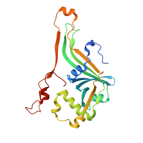
    4Y9I, 4YBN, 4ZKY, 5BNC

  • PubMed Abstract: 

    The deazaflavin cofactor F420 enhances the persistence of mycobacteria during hypoxia, oxidative stress, and antibiotic treatment. However, the identities and functions of the mycobacterial enzymes that utilize F420 under these conditions have yet to be resolved. In this work, we used sequence similarity networks to analyze the distribution of the largest F420-dependent protein family in mycobacteria. We show that these enzymes are part of a larger split β-barrel enzyme superfamily (flavin/deazaflavin oxidoreductases, FDORs) that include previously characterized pyridoxamine/pyridoxine-5'-phosphate oxidases and heme oxygenases. We show that these proteins variously utilize F420, flavin mononucleotide, flavin adenine dinucleotide, and heme cofactors. Functional annotation using phylogenetic, structural, and spectroscopic methods revealed their involvement in heme degradation, biliverdin reduction, fatty acid modification, and quinone reduction. Four novel crystal structures show that plasticity in substrate binding pockets and modifications to cofactor binding motifs enabled FDORs to carry out a variety of functions. This systematic classification and analysis provides a framework for further functional analysis of the roles of FDORs in mycobacterial pathogenesis and persistence.


  • Organizational Affiliation
    • Australian National University Research School of Chemistry, Sullivans Creek Road, Acton, ACT 2601, Australia.

Macromolecule Content 

  • Total Structure Weight: 50.75 kDa 
  • Atom Count: 3,805 
  • Modeled Residue Count: 419 
  • Deposited Residue Count: 448 
  • Unique protein chains: 1

Macromolecules

Find similar proteins by:|  3D Structure
Entity ID: 1
MoleculeChains  Sequence LengthOrganismDetailsImage
Flavin-nucleotide-binding protein
A, B
224Mycolicibacterium smegmatis MC2 155Mutation(s): 0 
Gene Names: MSMEG_4975MSMEI_4848LJ00_24605
UniProt
Find proteins for A0R238 (Mycolicibacterium smegmatis (strain ATCC 700084 / mc(2)155))
Explore A0R238 
Go to UniProtKB:  A0R238
Entity Groups
Sequence Clusters30% Identity50% Identity70% Identity90% Identity95% Identity100% Identity
UniProt GroupA0R238
Sequence Annotations
Expand
Reference Sequence

Small Molecules

Ligands 5 Unique
IDChains Name / Formula / InChI Key2D Diagram3D Interactions
FAD

Query on FAD


Download:Ideal Coordinates CCD File
H [auth B],
K [auth B]
FLAVIN-ADENINE DINUCLEOTIDE
C27 H33 N9 O15 P2
VWWQXMAJTJZDQX-UYBVJOGSSA-N
HEM

Query on HEM


Download:Ideal Coordinates CCD File
F [auth A],
G [auth B]
PROTOPORPHYRIN IX CONTAINING FE
C34 H32 Fe N4 O4
KABFMIBPWCXCRK-RGGAHWMASA-L
SO4

Query on SO4


Download:Ideal Coordinates CCD File
D [auth A],
J [auth B]
SULFATE ION
O4 S
QAOWNCQODCNURD-UHFFFAOYSA-L
ACT

Query on ACT


Download:Ideal Coordinates CCD File
E [auth A]ACETATE ION
C2 H3 O2
QTBSBXVTEAMEQO-UHFFFAOYSA-M
NI

Query on NI


Download:Ideal Coordinates CCD File
C [auth A],
I [auth B]
NICKEL (II) ION
Ni
VEQPNABPJHWNSG-UHFFFAOYSA-N

Experimental Data & Validation

Experimental Data

  • Method: X-RAY DIFFRACTION
  • Resolution: 1.90 Å
  • R-Value Free:  0.223 (Depositor), 0.228 (DCC) 
  • R-Value Work:  0.174 (Depositor), 0.185 (DCC) 
  • R-Value Observed: 0.176 (Depositor) 
Space Group: C 1 2 1
Unit Cell:
Length ( Å )Angle ( ˚ )
a = 84.722α = 90
b = 59.876β = 93.83
c = 89.658γ = 90
Software Package:
Software NamePurpose
REFMACrefinement
XDSdata reduction
Aimlessdata scaling
PHASERphasing
PHENIXrefinement

Structure Validation

View Full Validation Report


Ligand Structure Quality Assessment 


Entry History 

& Funding Information

Deposition Data


Funding OrganizationLocationGrant Number
Australian Research Council (ARC)Australia--

Revision History  (Full details and data files)

  • Version 1.0: 2015-10-21
    Type: Initial release
  • Version 1.1: 2015-11-04
    Changes: Database references, Derived calculations
  • Version 1.2: 2020-01-01
    Changes: Author supporting evidence, Data collection, Derived calculations
  • Version 1.3: 2023-09-27
    Changes: Data collection, Database references, Derived calculations, Refinement description