4YBF | pdb_00004ybf

Aspartic Proteinase Sapp2 Secreted from Candida Parapsilosis at 1.25 A Resolution


Experimental Data Snapshot

  • Method: X-RAY DIFFRACTION
  • Resolution: 1.24 Å
  • R-Value Free: 
    0.174 (Depositor), 0.187 (DCC) 
  • R-Value Work: 
    0.147 (Depositor) 
  • R-Value Observed: 
    0.148 (Depositor) 

Starting Model: experimental
View more details

wwPDB Validation 3D Report Full Report

Validation slider image for 4YBF

This is version 1.2 of the entry. See complete history

Literature

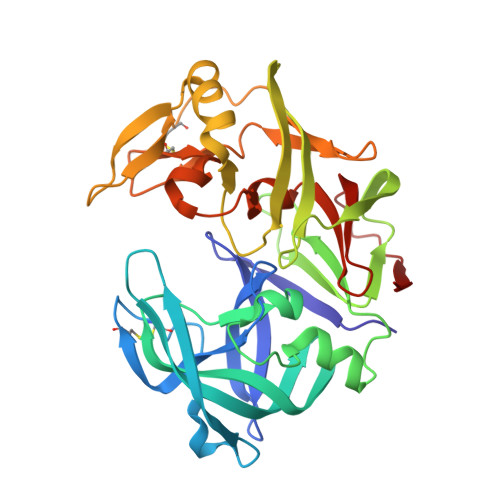
Atomic resolution crystal structure of Sapp2p, a secreted aspartic protease from Candida parapsilosis.

Dostal, J.Pecina, A.Hruskova-Heidingsfeldova, O.Mareckova, L.Pichova, I.Rezacova, P.Lepsik, M.Brynda, J.

(2015) Acta Crystallogr D Biol Crystallogr 71: 2494-2504

  • DOI: https://doi.org/10.1107/S1399004715019392
  • Primary Citation Related Structures: 
    4Y9W, 4YBF

  • PubMed Abstract: 

    The virulence of the Candida pathogens is enhanced by the production of secreted aspartic proteases, which therefore represent possible targets for drug design. Here, the crystal structure of the secreted aspartic protease Sapp2p from Candida parapsilosis was determined. Sapp2p was isolated from its natural source and crystallized in complex with pepstatin A, a classical aspartic protease inhibitor. The atomic resolution of 0.83 Å allowed the protonation states of the active-site residues to be inferred. A detailed comparison of the structure of Sapp2p with the structure of Sapp1p, the most abundant C. parapsilosis secreted aspartic protease, was performed. The analysis, which included advanced quantum-chemical interaction-energy calculations, uncovered molecular details that allowed the experimentally observed equipotent inhibition of both isoenzymes by pepstatin A to be rationalized.


  • Organizational Affiliation
    • Institute of Organic Chemistry and Biochemistry (IOCB), Academy of Sciences of the Czech Republic, Flemingovo náměstí 2, 166 10 Prague 6, Czech Republic.

Macromolecule Content 

  • Total Structure Weight: 35.58 kDa 
  • Atom Count: 2,852 
  • Modeled Residue Count: 334 
  • Deposited Residue Count: 334 
  • Unique protein chains: 1

Macromolecules

Find similar proteins by:|  3D Structure
Entity ID: 1
MoleculeChains  Sequence LengthOrganismDetailsImage
Candidapepsin-2334Candida parapsilosis CDC317Mutation(s): 0 
Gene Names: CPAR2_102580
EC: 3.4.23.24
UniProt
Find proteins for G8B6Y8 (Candida parapsilosis (strain CDC 317 / ATCC MYA-4646))
Explore G8B6Y8 
Go to UniProtKB:  G8B6Y8
Entity Groups
Sequence Clusters30% Identity50% Identity70% Identity90% Identity95% Identity100% Identity
UniProt GroupG8B6Y8
Sequence Annotations
Expand
Reference Sequence

Small Molecules

Ligands 1 Unique
IDChains Name / Formula / InChI Key2D Diagram3D Interactions
PEG

Query on PEG


Download:Ideal Coordinates CCD File
B [auth A]DI(HYDROXYETHYL)ETHER
C4 H10 O3
MTHSVFCYNBDYFN-UHFFFAOYSA-N

Experimental Data & Validation

Experimental Data

  • Method: X-RAY DIFFRACTION
  • Resolution: 1.24 Å
  • R-Value Free:  0.174 (Depositor), 0.187 (DCC) 
  • R-Value Work:  0.147 (Depositor) 
  • R-Value Observed: 0.148 (Depositor) 
Space Group: P 1 21 1
Unit Cell:
Length ( Å )Angle ( ˚ )
a = 49.138α = 90
b = 57.147β = 90.61
c = 54.553γ = 90
Software Package:
Software NamePurpose
XSCALEdata scaling
REFMACrefinement
PDB_EXTRACTdata extraction
XDSdata reduction
MOLREPphasing

Structure Validation

View Full Validation Report


Entry History 

Deposition Data

Revision History  (Full details and data files)

  • Version 1.0: 2016-01-27
    Type: Initial release
  • Version 1.1: 2024-01-10
    Changes: Data collection, Database references, Refinement description
  • Version 1.2: 2024-11-20
    Changes: Structure summary