4Y97 | pdb_00004y97

Crystal Structure of human Pol alpha B-subunit in complex with C-terminal domain of catalytic subunit


Experimental Data Snapshot

  • Method: X-RAY DIFFRACTION
  • Resolution: 2.51 Å
  • R-Value Free: 
    0.257 (Depositor), 0.264 (DCC) 
  • R-Value Work: 
    0.218 (Depositor), 0.228 (DCC) 
  • R-Value Observed: 
    0.218 (Depositor) 

Starting Model: experimental
View more details

wwPDB Validation 3D Report Full Report

Validation slider image for 4Y97

This is version 1.5 of the entry. See complete history

Literature

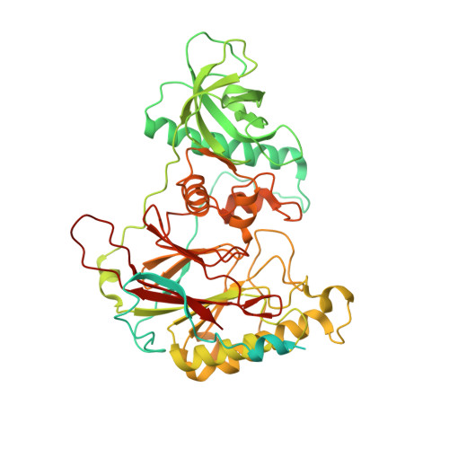
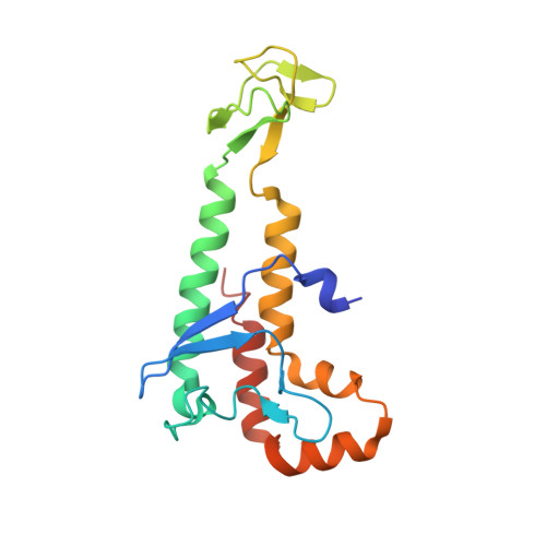
Crystal Structure of the Human Pol alpha B Subunit in Complex with the C-terminal Domain of the Catalytic Subunit.

Suwa, Y.Gu, J.Baranovskiy, A.G.Babayeva, N.D.Pavlov, Y.I.Tahirov, T.H.

(2015) J Biological Chem 290: 14328-14337

  • DOI: https://doi.org/10.1074/jbc.M115.649954
  • Primary Citation Related Structures: 
    4Y97

  • PubMed Abstract: 

    In eukaryotic DNA replication, short RNA-DNA hybrid primers synthesized by primase-DNA polymerase α (Prim-Pol α) are needed to start DNA replication by the replicative DNA polymerases, Pol δ and Pol ϵ. The C terminus of the Pol α catalytic subunit (p180C) in complex with the B subunit (p70) regulates the RNA priming and DNA polymerizing activities of Prim-Pol α. It tethers Pol α and primase, facilitating RNA primer handover from primase to Pol α. To understand these regulatory mechanisms and to reveal the details of human Pol α organization, we determined the crystal structure of p70 in complex with p180C. The structured portion of p70 includes a phosphodiesterase (PDE) domain and an oligonucleotide/oligosaccharide binding (OB) domain. The N-terminal domain and the linker connecting it to the PDE domain are disordered in the reported crystal structure. The p180C adopts an elongated asymmetric saddle shape, with a three-helix bundle in the middle and zinc-binding modules (Zn1 and Zn2) on each side. The extensive p180C-p70 interactions involve 20 hydrogen bonds and a number of hydrophobic interactions resulting in an extended buried surface of 4080 Å(2). Importantly, in the structure of the p180C-p70 complex with full-length p70, the residues from the N-terminal to the OB domain contribute to interactions with p180C. The comparative structural analysis revealed both the conserved features and the differences between the human and yeast Pol α complexes.


  • Organizational Affiliation
    • From the Eppley Institute for Research in Cancer and Allied Diseases and.

Macromolecule Content 

  • Total Structure Weight: 349.35 kDa 
  • Atom Count: 19,724 
  • Modeled Residue Count: 2,464 
  • Deposited Residue Count: 3,112 
  • Unique protein chains: 2

Macromolecules

Find similar proteins by:|  3D Structure
Entity ID: 1
MoleculeChains  Sequence LengthOrganismDetailsImage
DNA polymerase alpha subunit B
A, C, E, G
598Homo sapiensMutation(s): 0 
Gene Names: POLA2
UniProt & NIH Common Fund Data Resources
Find proteins for Q14181 (Homo sapiens)
Explore Q14181 
Go to UniProtKB:  Q14181
PHAROS:  Q14181
GTEx:  ENSG00000014138 
Entity Groups
Sequence Clusters30% Identity50% Identity70% Identity90% Identity95% Identity100% Identity
UniProt GroupQ14181
Sequence Annotations
Expand
Reference Sequence
Find similar proteins by:|  3D Structure
Entity ID: 2
MoleculeChains  Sequence LengthOrganismDetailsImage
DNA polymerase alpha catalytic subunit
B, D, F, H
180Homo sapiensMutation(s): 0 
Gene Names: POLA1POLA
EC: 2.7.7.7
UniProt & NIH Common Fund Data Resources
Find proteins for P09884 (Homo sapiens)
Explore P09884 
Go to UniProtKB:  P09884
PHAROS:  P09884
GTEx:  ENSG00000101868 
Entity Groups
Sequence Clusters30% Identity50% Identity70% Identity90% Identity95% Identity100% Identity
UniProt GroupP09884
Sequence Annotations
Expand
Reference Sequence

Experimental Data & Validation

Experimental Data

  • Method: X-RAY DIFFRACTION
  • Resolution: 2.51 Å
  • R-Value Free:  0.257 (Depositor), 0.264 (DCC) 
  • R-Value Work:  0.218 (Depositor), 0.228 (DCC) 
  • R-Value Observed: 0.218 (Depositor) 
Space Group: P 1 21 1
Unit Cell:
Length ( Å )Angle ( ˚ )
a = 103.593α = 90
b = 137.135β = 100.97
c = 132.092γ = 90
Software Package:
Software NamePurpose
CNSrefinement
HKL-2000data reduction
HKL-2000data scaling
PHENIXphasing

Structure Validation

View Full Validation Report


Entry History 

& Funding Information

Deposition Data


Funding OrganizationLocationGrant Number
National Institutes of Health/National Institute of General Medical Sciences (NIH/NIGMS)United StatesGM101167

Revision History  (Full details and data files)

  • Version 1.0: 2015-04-15
    Type: Initial release
  • Version 1.1: 2015-04-22
    Changes: Database references
  • Version 1.2: 2015-06-17
    Changes: Database references
  • Version 1.3: 2017-09-13
    Changes: Author supporting evidence, Database references, Derived calculations, Source and taxonomy
  • Version 1.4: 2019-12-25
    Changes: Author supporting evidence
  • Version 1.5: 2023-09-27
    Changes: Data collection, Database references, Refinement description