4Y5Z | pdb_00004y5z

T=1 capsid structure of SeMV Ndel65CP fused with B-domain of S. aureus protein SpA at the N-terminus (P1 crystal form)


Experimental Data Snapshot

  • Method: X-RAY DIFFRACTION
  • Resolution: 2.95 Å
  • R-Value Free: 
    0.249 (Depositor), 0.253 (DCC) 
  • R-Value Work: 
    0.196 (Depositor), 0.203 (DCC) 
  • R-Value Observed: 
    0.199 (Depositor) 

Starting Model: experimental
View more details

wwPDB Validation 3D Report Full Report

Validation slider image for 4Y5Z

This is version 1.3 of the entry. See complete history

Literature

Structural studies on chimeric Sesbania mosaic virus coat protein: Revisiting SeMV assembly.

Gulati, A.Murthy, A.Abraham, A.Mohan, K.Natraj, U.Savithri, H.S.Murthy, M.R.

(2015) Virology 489: 34-43

  • DOI: https://doi.org/10.1016/j.virol.2015.11.029
  • Primary Citation Related Structures: 
    4Y4Y, 4Y5Z

  • PubMed Abstract: 

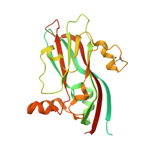
    The capsid protein (CP) of Sesbania mosaic virus (SeMV, a T=3 plant virus) consists of a disordered N-terminal R-domain and an ordered S-domain. Removal of the R-domain results in the formation of T=1 particles. In the current study, the R-domain was replaced with unrelated polypeptides of similar lengths: the B-domain of Staphylococcus aureus SpA, and SeMV encoded polypeptides P8 and P10. The chimeric proteins contained T=3 or larger virus-like particles (VLPs) and could not be crystallized. The presence of metal ions during purification resulted in a large number of heterogeneous nucleoprotein complexes. N∆65-B (R domain replaced with B domain) could also be purified in a dimeric form. Its crystal structure revealed T=1 particles devoid of metal ions and the B-domain was disordered. However, the B-domain was functional in N∆65-B VLPs, suggesting possible biotechnological applications. These studies illustrate the importance of N-terminal residues, metal ions and robustness of the assembly process.


  • Organizational Affiliation
    • Molecular Biophysics Unit, Indian Institute of Science, Bangalore 560012, India.

Macromolecule Content 

  • Total Structure Weight: 1,833.75 kDa 
  • Atom Count: 86,922 
  • Modeled Residue Count: 11,534 
  • Deposited Residue Count: 16,920 
  • Unique protein chains: 1

Macromolecules

Find similar proteins by:|  3D Structure
Entity ID: 1
MoleculeChains  Sequence LengthOrganismDetailsImage
Immunoglobulin G-binding protein A,Coat protein282Staphylococcus aureus subsp. aureus NCTC 8325Sesbania mosaic virus
This entity is chimeric
Mutation(s): 0 
Gene Names: spaSAOUHSC_00069
UniProt
Find proteins for P02976 (Staphylococcus aureus (strain NCTC 8325 / PS 47))
Explore P02976 
Go to UniProtKB:  P02976
Find proteins for P38507 (Staphylococcus aureus)
Go to UniProtKB:  P38507
Find proteins for Q9EB06 (Sesbania mosaic virus)
Explore Q9EB06 
Go to UniProtKB:  Q9EB06
Entity Groups
Sequence Clusters30% Identity50% Identity70% Identity90% Identity95% Identity100% Identity
UniProt GroupsP02976Q9EB06
Sequence Annotations
Expand
Reference Sequence

Small Molecules

Ligands 1 Unique
IDChains Name / Formula / InChI Key2D Diagram3D Interactions
SO4

Query on SO4


Download:Ideal Coordinates CCD File
IB [auth A]
JB [auth H]
KB [auth M]
LB [auth P]
MB [auth X]
IB [auth A],
JB [auth H],
KB [auth M],
LB [auth P],
MB [auth X],
NB [auth Z],
OB [auth o],
PB [auth s],
QB [auth w],
RB [auth 4],
SB [auth 7],
TB [auth h]
SULFATE ION
O4 S
QAOWNCQODCNURD-UHFFFAOYSA-L

Experimental Data & Validation

Experimental Data

  • Method: X-RAY DIFFRACTION
  • Resolution: 2.95 Å
  • R-Value Free:  0.249 (Depositor), 0.253 (DCC) 
  • R-Value Work:  0.196 (Depositor), 0.203 (DCC) 
  • R-Value Observed: 0.199 (Depositor) 
Space Group: P 1
Unit Cell:
Length ( Å )Angle ( ˚ )
a = 186.48α = 61.09
b = 187.42β = 89.24
c = 187.93γ = 60.2
Software Package:
Software NamePurpose
SCALAdata scaling
PHENIXrefinement
PDB_EXTRACTdata extraction
MOSFLMdata reduction
PHASERphasing

Structure Validation

View Full Validation Report


Entry History 

& Funding Information

Deposition Data


Funding OrganizationLocationGrant Number
DST, DBT, J.C BoseIndia--

Revision History  (Full details and data files)

  • Version 1.0: 2016-01-20
    Type: Initial release
  • Version 1.1: 2023-11-08
    Changes: Data collection, Database references, Derived calculations, Refinement description
  • Version 1.2: 2023-11-29
    Changes: Database references
  • Version 1.3: 2024-11-06
    Changes: Structure summary