4Y5D | pdb_00004y5d

CRYSTAL STRUCTURE OF ALiS2-STREPTAVIDIN COMPLEX


Experimental Data Snapshot

  • Method: X-RAY DIFFRACTION
  • Resolution: 1.20 Å
  • R-Value Free: 
    0.174 (Depositor), 0.174 (DCC) 
  • R-Value Work: 
    0.138 (Depositor), 0.139 (DCC) 
  • R-Value Observed: 
    0.140 (Depositor) 

wwPDB Validation 3D Report Full Report

Validation slider image for 4Y5D

Ligand Structure Quality Assessment 


This is version 1.3 of the entry. See complete history

Literature

Artificial Ligands of Streptavidin (ALiS): Discovery, Characterization, and Application for Reversible Control of Intracellular Protein Transport

Terai, T.Kohno, M.Boncompain, G.Sugiyama, S.Saito, N.Fujikake, R.Ueno, T.Komatsu, T.Hanaoka, K.Okabe, T.Urano, Y.Perez, F.Nagano, T.

(2015) J Am Chem Soc 137: 10464-10467

  • DOI: https://doi.org/10.1021/jacs.5b05672
  • Primary Citation Related Structures: 
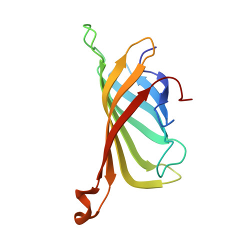
    4Y59, 4Y5D

  • PubMed Abstract: 

    Artificial ligands of streptavidin (ALiS) with association constants of ∼10(6) M(-1) were discovered by high-throughput screening of our chemical library, and their binding characteristics, including X-ray crystal structure of the streptavidin complex, were determined. Unlike biotin and its derivatives, ALiS exhibits fast dissociation kinetics and excellent cell permeability. The streptavidin-ALiS system provides a novel, practical compound-dependent methodology for repeated reversible cycling of protein localization between intracellular organella.


  • Organizational Affiliation
    • Graduate School of Science, Osaka University , 2-1 Yamadaoka, Suita, Osaka 565-0871, Japan.

Macromolecule Content 

  • Total Structure Weight: 53.49 kDa 
  • Atom Count: 4,380 
  • Modeled Residue Count: 488 
  • Deposited Residue Count: 488 
  • Unique protein chains: 1

Macromolecules

Find similar proteins by:|  3D Structure
Entity ID: 1
MoleculeChains  Sequence LengthOrganismDetailsImage
Streptavidin
A, B, C, D
122Streptomyces avidiniiMutation(s): 0 
UniProt
Find proteins for P22629 (Streptomyces avidinii)
Explore P22629 
Go to UniProtKB:  P22629
Entity Groups
Sequence Clusters30% Identity50% Identity70% Identity90% Identity95% Identity100% Identity
UniProt GroupP22629
Sequence Annotations
Expand
Reference Sequence

Small Molecules

Ligands 4 Unique
IDChains Name / Formula / InChI Key2D Diagram3D Interactions
PE3

Query on PE3


Download:Ideal Coordinates CCD File
F [auth A]3,6,9,12,15,18,21,24,27,30,33,36,39-TRIDECAOXAHENTETRACONTANE-1,41-DIOL
C28 H58 O15
ILLKMACMBHTSHP-UHFFFAOYSA-N
P6G

Query on P6G


Download:Ideal Coordinates CCD File
I [auth C],
J [auth C]
HEXAETHYLENE GLYCOL
C12 H26 O7
IIRDTKBZINWQAW-UHFFFAOYSA-N
MT6

Query on MT6


Download:Ideal Coordinates CCD File
E [auth A],
G [auth B],
K [auth D]
methyl 3-(4-oxo-4,5-dihydrofuro[3,2-c]pyridin-2-yl)benzoate
C15 H11 N O4
OVTGPKXYLOQTEQ-UHFFFAOYSA-N
DMS

Query on DMS


Download:Ideal Coordinates CCD File
H [auth C]DIMETHYL SULFOXIDE
C2 H6 O S
IAZDPXIOMUYVGZ-UHFFFAOYSA-N

Experimental Data & Validation

Experimental Data

  • Method: X-RAY DIFFRACTION
  • Resolution: 1.20 Å
  • R-Value Free:  0.174 (Depositor), 0.174 (DCC) 
  • R-Value Work:  0.138 (Depositor), 0.139 (DCC) 
  • R-Value Observed: 0.140 (Depositor) 
Space Group: P 1 21 1
Unit Cell:
Length ( Å )Angle ( ˚ )
a = 57.927α = 90
b = 83.774β = 98.75
c = 45.947γ = 90
Software Package:
Software NamePurpose
REFMACrefinement
HKL-2000data reduction
HKL-2000data scaling
MOLREPphasing

Structure Validation

View Full Validation Report


Ligand Structure Quality Assessment 


Entry History 

Deposition Data

Revision History  (Full details and data files)

  • Version 1.0: 2015-09-23
    Type: Initial release
  • Version 1.1: 2015-12-16
    Changes: Database references
  • Version 1.2: 2020-02-05
    Changes: Data collection, Derived calculations
  • Version 1.3: 2024-03-20
    Changes: Data collection, Database references