4Y42 | pdb_00004y42

Cyanase (CynS) from Serratia proteamaculans


Experimental Data Snapshot

  • Method: X-RAY DIFFRACTION
  • Resolution: 2.09 Å
  • R-Value Free: 
    0.200 (Depositor), 0.199 (DCC) 
  • R-Value Work: 
    0.150 (Depositor), 0.149 (DCC) 
  • R-Value Observed: 
    0.152 (Depositor) 

Starting Model: experimental
View more details

wwPDB Validation 3D Report Full Report

Validation slider image for 4Y42

This is version 1.2 of the entry. See complete history

Literature

Serendipitous crystallization and structure determination of cyanase (CynS) from Serratia proteamaculans

Butryn, A.Stoehr, G.Linke-Winnebeck, C.Hopfner, K.-P.

(2015) Acta Crystallogr F Struct Biol Commun 71: 471-476

  • DOI: https://doi.org/10.1107/S2053230X15004902
  • Primary Citation Related Structures: 
    4Y42

  • PubMed Abstract: 

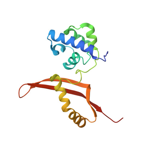
    Cyanate hydratase (CynS) catalyzes the decomposition of cyanate and bicarbonate into ammonia and carbon dioxide. Here, the serendipitous crystallization of CynS from Serratia proteamaculans (SpCynS) is reported. SpCynS was crystallized as an impurity and its identity was determined using mass-spectrometric analysis. The crystals belonged to space group P1 and diffracted to 2.1 Å resolution. The overall structure of SpCynS is very similar to a previously determined structure of CynS from Escherichia coli. Density for a ligand bound to the SpCynS active site was observed, but could not be unambiguously identified. Additionally, glycerol molecules bound at the entry to the active site of the enzyme indicate conserved residues that might be important for the trafficking of substrates and products.


  • Organizational Affiliation
    • Gene Center and Department of Biochemistry, Ludwig-Maximilians-University Munich, Feodor-Lynen-Strasse 25, 81377 Munich, Germany.

Macromolecule Content 

  • Total Structure Weight: 172.1 kDa 
  • Atom Count: 13,413 
  • Modeled Residue Count: 1,550 
  • Deposited Residue Count: 1,560 
  • Unique protein chains: 1

Macromolecules

Find similar proteins by:|  3D Structure
Entity ID: 1
MoleculeChains  Sequence LengthOrganismDetailsImage
Cyanate hydratase
A, B, C, D, E
A, B, C, D, E, F, G, H, I, J
156Serratia proteamaculans 568Mutation(s): 0 
EC: 4.2.1.104
UniProt
Find proteins for A8GBZ7 (Serratia proteamaculans (strain 568))
Explore A8GBZ7 
Go to UniProtKB:  A8GBZ7
Entity Groups
Sequence Clusters30% Identity50% Identity70% Identity90% Identity95% Identity100% Identity
UniProt GroupA8GBZ7
Sequence Annotations
Expand
Reference Sequence

Experimental Data & Validation

Experimental Data

  • Method: X-RAY DIFFRACTION
  • Resolution: 2.09 Å
  • R-Value Free:  0.200 (Depositor), 0.199 (DCC) 
  • R-Value Work:  0.150 (Depositor), 0.149 (DCC) 
  • R-Value Observed: 0.152 (Depositor) 
Space Group: P 1
Unit Cell:
Length ( Å )Angle ( ˚ )
a = 82.549α = 98.56
b = 82.456β = 117.38
c = 87.985γ = 110.46
Software Package:
Software NamePurpose
PHENIXrefinement
XDSdata reduction
XSCALEdata scaling
PHASERphasing

Structure Validation

View Full Validation Report


Entry History 

Deposition Data

Revision History  (Full details and data files)

  • Version 1.0: 2015-04-08
    Type: Initial release
  • Version 1.1: 2015-04-22
    Changes: Database references
  • Version 1.2: 2024-01-10
    Changes: Data collection, Database references, Refinement description