4XZM | pdb_00004xzm

Crystal structure of the methylated wild-type AKR1B10 holoenzyme


Experimental Data Snapshot

  • Method: X-RAY DIFFRACTION
  • Resolution: 1.75 Å
  • R-Value Free: 
    0.250 (Depositor), 0.250 (DCC) 
  • R-Value Work: 
    0.203 (Depositor), 0.201 (DCC) 
  • R-Value Observed: 
    0.205 (Depositor) 

Starting Model: experimental
View more details

wwPDB Validation 3D Report Full Report

Validation slider image for 4XZM

Ligand Structure Quality Assessment 


This is version 1.2 of the entry. See complete history

Literature

Structural Determinants of the Selectivity of 3-Benzyluracil-1-acetic Acids toward Human Enzymes Aldose Reductase and AKR1B10.

Ruiz, F.X.Cousido-Siah, A.Porte, S.Dominguez, M.Crespo, I.Rechlin, C.Mitschler, A.de Lera, A.R.Martin, M.J.de la Fuente, J.A.Klebe, G.Pares, X.Farres, J.Podjarny, A.

(2015) ChemMedChem 10: 1989-2003

  • DOI: https://doi.org/10.1002/cmdc.201500393
  • Primary Citation Related Structures: 
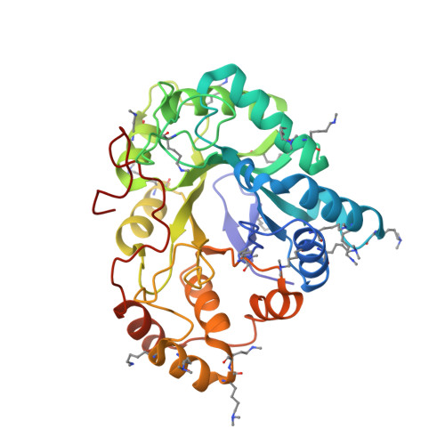
    4XZH, 4XZI, 4XZL, 4XZM, 4XZN

  • PubMed Abstract: 

    The human enzymes aldose reductase (AR) and AKR1B10 have been thoroughly explored in terms of their roles in diabetes, inflammatory disorders, and cancer. In this study we identified two new lead compounds, 2-(3-(4-chloro-3-nitrobenzyl)-2,4-dioxo-3,4-dihydropyrimidin-1(2H)-yl)acetic acid (JF0048, 3) and 2-(2,4-dioxo-3-(2,3,4,5-tetrabromo-6-methoxybenzyl)-3,4-dihydropyrimidin-1(2H)-yl)acetic acid (JF0049, 4), which selectively target these enzymes. Although 3 and 4 share the 3-benzyluracil-1-acetic acid scaffold, they have different substituents in their aryl moieties. Inhibition studies along with thermodynamic and structural characterizations of both enzymes revealed that the chloronitrobenzyl moiety of compound 3 can open the AR specificity pocket but not that of the AKR1B10 cognate. In contrast, the larger atoms at the ortho and/or meta positions of compound 4 prevent the AR specificity pocket from opening due to steric hindrance and provide a tighter fit to the AKR1B10 inhibitor binding pocket, probably enhanced by the displacement of a disordered water molecule trapped in a hydrophobic subpocket, creating an enthalpic signature. Furthermore, this selectivity also occurs in the cell, which enables the development of a more efficient drug design strategy: compound 3 prevents sorbitol accumulation in human retinal ARPE-19 cells, whereas 4 stops proliferation in human lung cancer NCI-H460 cells.


  • Organizational Affiliation
    • Department of Integrative Biology, Institut de Génétique et de Biologie Moléculaire et Cellulaire, CNRS, INSERM, UdS, rue Laurent Fries, 67404, Illkirch CEDEX, France. fxavier.ruiz@gmail.com.

Macromolecule Content 

  • Total Structure Weight: 37.22 kDa 
  • Atom Count: 2,846 
  • Modeled Residue Count: 316 
  • Deposited Residue Count: 316 
  • Unique protein chains: 1

Macromolecules

Find similar proteins by:|  3D Structure
Entity ID: 1
MoleculeChains  Sequence LengthOrganismDetailsImage
Aldo-keto reductase family 1 member B10A [auth X]316Homo sapiensMutation(s): 0 
Gene Names: AKR1B10AKR1B11
EC: 1.1.1 (PDB Primary Data), 1.1.1.300 (UniProt), 1.1.1.54 (UniProt)
UniProt & NIH Common Fund Data Resources
Find proteins for O60218 (Homo sapiens)
Explore O60218 
Go to UniProtKB:  O60218
PHAROS:  O60218
GTEx:  ENSG00000198074 
Entity Groups
Sequence Clusters30% Identity50% Identity70% Identity90% Identity95% Identity100% Identity
UniProt GroupO60218
Sequence Annotations
Expand
Reference Sequence

Small Molecules

Ligands 2 Unique
IDChains Name / Formula / InChI Key2D Diagram3D Interactions
NAP

Query on NAP


Download:Ideal Coordinates CCD File
B [auth X]NADP NICOTINAMIDE-ADENINE-DINUCLEOTIDE PHOSPHATE
C21 H28 N7 O17 P3
XJLXINKUBYWONI-NNYOXOHSSA-N
EDO

Query on EDO


Download:Ideal Coordinates CCD File
C [auth X]1,2-ETHANEDIOL
C2 H6 O2
LYCAIKOWRPUZTN-UHFFFAOYSA-N
Modified Residues  2 Unique
IDChains TypeFormula2D DiagramParent
MLY
Query on MLY
A [auth X]L-PEPTIDE LINKINGC8 H18 N2 O2LYS
MLZ
Query on MLZ
A [auth X]L-PEPTIDE LINKINGC7 H16 N2 O2LYS

Experimental Data & Validation

Experimental Data

  • Method: X-RAY DIFFRACTION
  • Resolution: 1.75 Å
  • R-Value Free:  0.250 (Depositor), 0.250 (DCC) 
  • R-Value Work:  0.203 (Depositor), 0.201 (DCC) 
  • R-Value Observed: 0.205 (Depositor) 
Space Group: P 31
Unit Cell:
Length ( Å )Angle ( ˚ )
a = 79.435α = 90
b = 79.435β = 90
c = 49.796γ = 120
Software Package:
Software NamePurpose
HKL-2000data reduction
HKL-2000data scaling
PHASERphasing
PHENIXrefinement

Structure Validation

View Full Validation Report


Ligand Structure Quality Assessment 


Entry History 

Deposition Data

Revision History  (Full details and data files)

  • Version 1.0: 2015-11-18
    Type: Initial release
  • Version 1.1: 2016-01-27
    Changes: Database references
  • Version 1.2: 2024-01-10
    Changes: Data collection, Database references, Refinement description